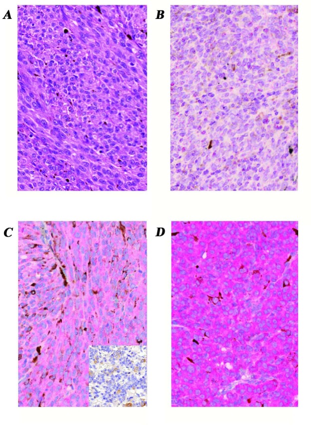
Figure 2. Expression of LHRH receptor protein in enucleated human uveal melanoma tissue samples demonstrated by immunoperoxidase staining.

A, Hematoxylin-eosin stained section of a representative sample shows melanin-producing neoplastic cells with spindle and epithelioid pattern. a-d, images of representative samples immunostained for type I LHRH receptor; B, tumor sample without detectable mRNA for type I LHRH receptor also revealed no identifiable LHRH receptor protein using IHC staining; C, representative tissue section shows mild positivity for type I LHRH receptor (faint red cytoplasms of tumor cells). Insert is a negative control for the staining-specificity (see Methods); D, representative tumor sample exhibits intense expression of type I LHRH receptor in nearly 100% of tumor cells (intense red cytoplasmic staining) which correlated with the corresponding mRNA levels. Original magnifications of all images: 40x. Images B-D are immunoperoxidase stained sections with hematoxylin nuclear counterstaining.
