Figure 5.
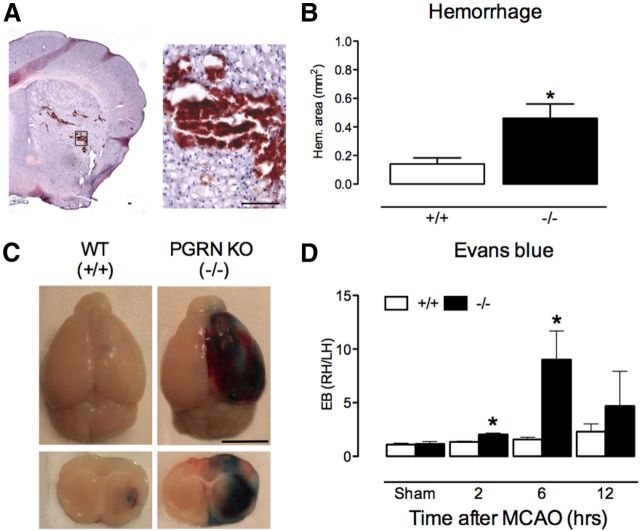
Post-ischemic hemorrhage and BBB disruption are enhanced in PGRN-KO. A, Representative H&E-stained brain section illustrating hemorrhage 6 h after MCAO in PGRN-KO mice. Early tissue injury is also seen as pallor in the dorsal striatum. Scale bar, 100 μm. B, The area of post-ischemic hemorrhage is greater in PGRN-KO (−/−) mice 6 h after MCAO compared with WT (+/+) mice (n = 8 per group; *p < 0.05, t test) C, Representative images of EB extravasation in WT and PGRN-KO mice 6 h after MCAO. Notice the increased BBB permeability in PGRN-KO mice. Scale bar, 5 mm. D, Temporal profile of EB extravasation after MCAO, expressed as ratio between ischemic and non-ischemic hemisphere, showing greater BBB permeability in PGRN-KO (n = 7–10 per group; *p < 0.05 from respective WT, t test).

