
Figure 2.
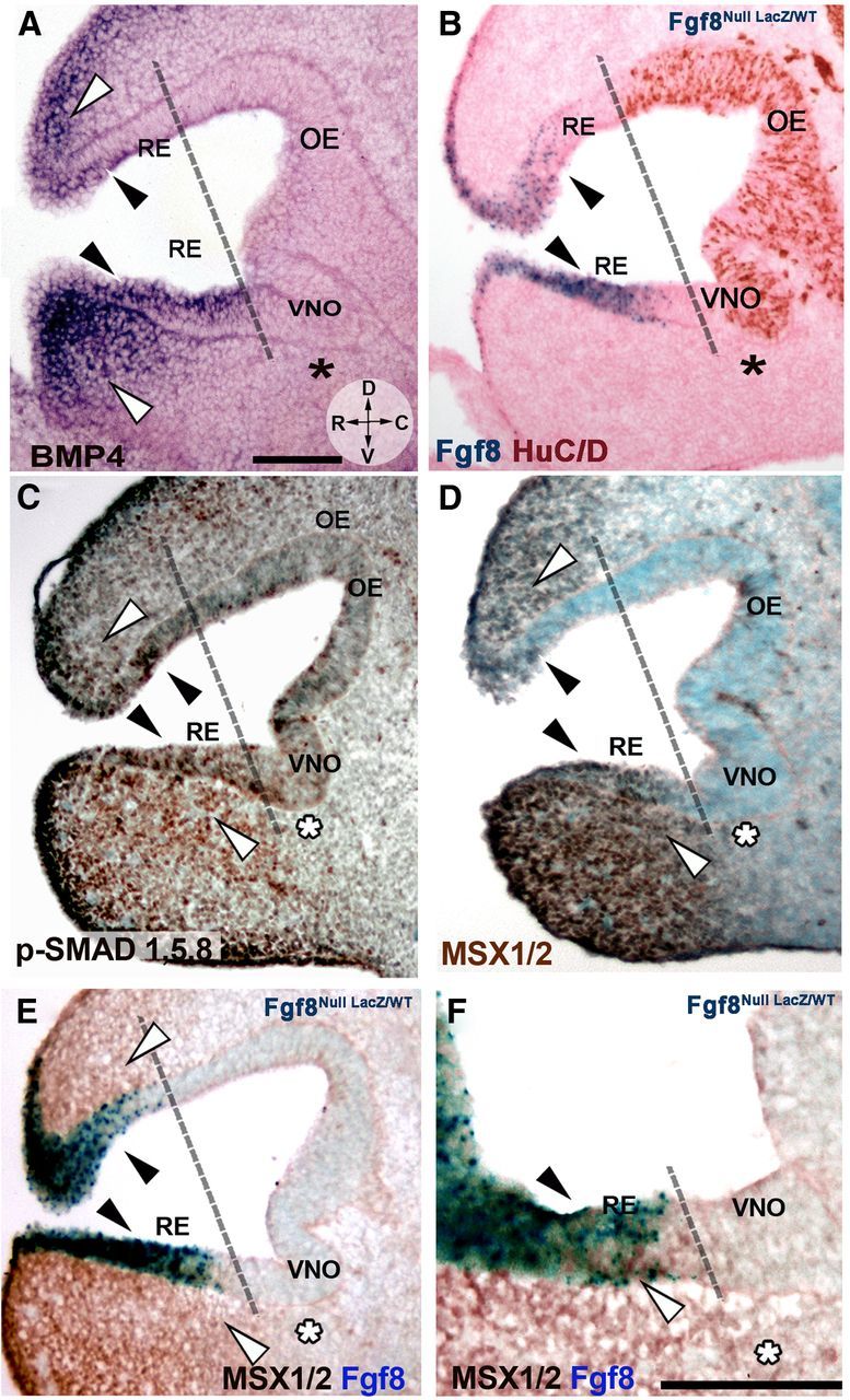
Bmp4 is expressed in the developing nasal mesenchyme and is coexpressed with FGF8 in the RE. A–D, E11.5, parasagittal sections, orientation indicated in A. R, Rostral; D, dorsal; V, ventral; C, caudal. Use dashed line as reference to separate non-neurogenic and neurogenic OP areas. A, Bmp4 in situ hybridization showed expression along the RE (black arrowheads) and in rostral, dorsal, and ventral nasal mesenchyme (white arrowheads). Bmp4 expression was not detected in the caudal nasal mesenchyme (asterisk) facing the neurogenic VNO (compare to B). B, Fgf8nullLacz/WT; X-gal enzymatic reaction (blue) and HuC/D immunostaining (brown) highlights the occurrence of neurogenesis mainly caudal to Fgf8-expressing areas (blue). Fgf8 was expressed in areas where Bmp4 expression was detected (compare black arrows in A and B). C, Immunostaining for p-SMAD 1,5,8 (brown) detected active BMP4 downstream signaling in areas of Bmp4 expression (A, C, white arrowheads) and along the RE (black arrowheads). Decreased p-SMAD 1,5,8 immunoreactivity was detected in area facing the VNO (asterisk). D, MSX1/2 immunolabeling (dark brown) revealed a pattern similar to p-SMAD (compare with C) with high MSX1/2 levels in the RE (black arrowheads) and mesenchyme proximal to Bmp4 sources (white arrowheads; compare with A). No expression was found in the mesenchyme facing the neurogenic VNO (asterisk). E, F, MSX1/2 immunolabeling (brown) and Fgf8 expression (X-gal enzymatic reaction, blue) revealed co-localization in the RE (black arrowheads). Scale bars: (in A) A–D, 100 μm; F, 100 μm.