
Figure 5.
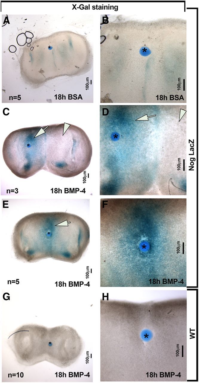
BMP4 directly induced Nog expression in developing nasal mesenchyme. A–F, Coronal nasal explant from NognullLacz/WT. A, B, Control bead soaked in BSA did not induce Nog expression in the surrounding tissue. C–F, BMP4-soaked bead placed on lateral (C, D) or midline (E, F) mesenchymal tissue of explants from Nog-LacZ mice induced Nog expression. Evidence of BMP4-induced expression of Nog was present regardless of bead position (see arrows in C, D, E vs internal negative control arrowheads in C, D). Endogenous levels of Nog served as internal controls in these experiments. BSA-soaked beads placed on Nog-LacZ explant tissue (A, B) as well as BMP4-soaked beads placed on explants from WT mice (G, H) served as negative controls and showed no induction of Nog:β-gal after X-gal staining. Bead is marked by asterisk in all images. High-magnification images are shown in B–H.