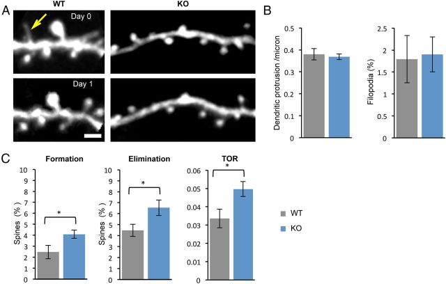
Figure 5.
Normal density but higher turnover rate of dendritic spines in the fmr1 KO mouse. A, Multiphoton in vivo imaging of dendritic spines in layer 1 of Thy1 YFP-H mice crossbred with fmr1 KO mice. Imaging was performed on two consecutive days in untrained mice. Yellow arrow points to a filopodia that was lost on the second day of imaging. Scale bar, 3 μm. B, Density of dendritic protrusion and percentage of filopodia were similar between the genotypes. C, Spine dynamics, measured as rates of spine formation, elimination, and TOR were higher in the KO mouse. WT (n = 7) and KO (n = 8) mice. Mean ± SEM. *p < 0.05.

