Figure 1.

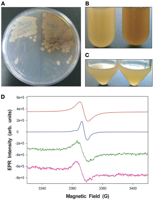
Pigmentation differences between V. campbellii WT and ΔhmgA. (A) Colonies grown on LM agar. WT (left), ΔhmgA (right); (B) Cell cultures grown in LM in borosilicate glass tubes (48 h). WT (left), ΔhmgA (right); (C) Cell cultures grown in LM in polypropylene conical tubes (48 h). WT (left), ΔhmgA (right); (D) EPR spectra of synthetic melanin (red), DHN-melanin (blue) and partially purified pigments from the supernatants of two independent ΔhmgA cultures (green and pink).
