
Figure 2.
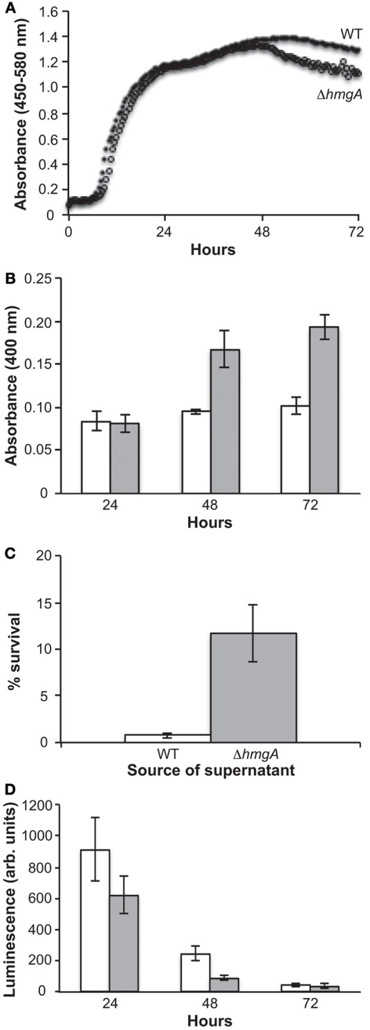
Growth, pigment production and bioluminescence in V. campbellii WT and ΔhmgA. (A) Grow curves of the WT and ΔhmgA strains in LM at 30°C. WT (closed circle), ΔhmgA (open circle); (B) Absorbance of pigment-containing supernatants. WT (white bars), ΔhmgA (gray bars); (C) Survival of WT V. campbellii cells in 2 mM H2O2 when resuspended in supernatants from WT or ΔhmgA cultures; (D) Bioluminescence measurements. WT (white bars), ΔhmgA (gray bars). Error bars represent the standard deviation from three experiments.