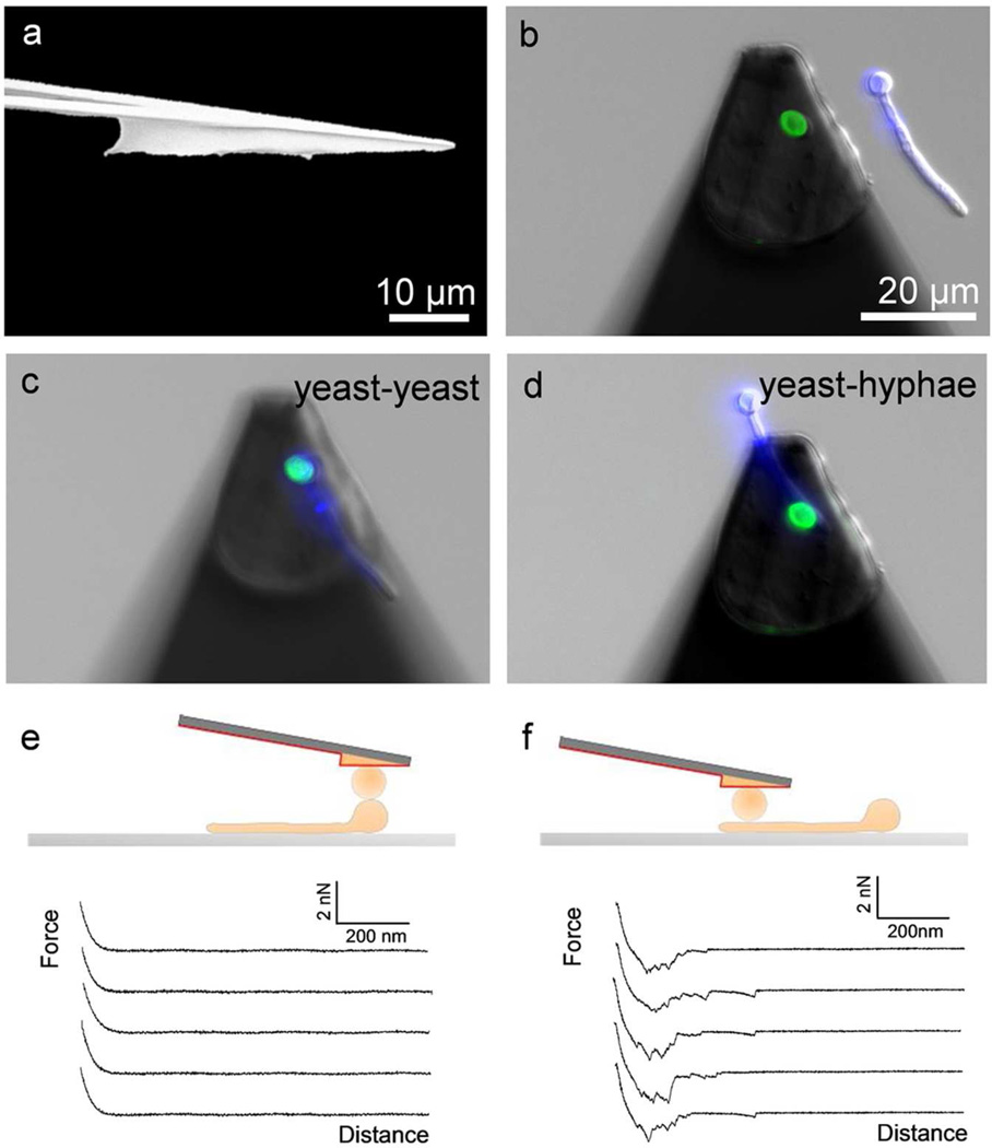
Figure 1.
Force spectroscopy of cell-cell adhesion using wedged cantilevers. (a) Scanning electron microscopy image of a wedged cantilever prepared using UV-curable glue. (b) Non-destructive method for studying yeast-hyphae adhesion: single yeast cells from Candida albicans (labeled with Con A-FITC, green) were attached on a polydopamine-coated wedged cantilever and approached towards a C. albicans hyphae (Calcofluor White, blue) immobilized on a hydrophobic substrate. (c-f) To measure yeast-hyphae adhesion forces, the yeast cell probe was positioned on top of the yeast region (c, e) or the germ tube region (d, f) of the hyphae, and multiple force-distance curves were recorded (e, f).

