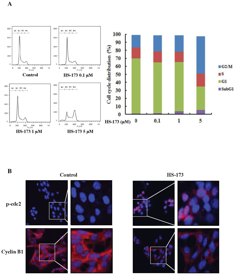
Figure 2. Effect of HS-173 on the HSC cell cycle.
(A) After incubating for 1 day, HSC-T6 cells were treated with various concentrations of HS-173 (0, 0.1, 1, and 5 μM) for 8 h, stained with propidium iodide (PI) and analyzed with a FACSCalibur flow cytometer. M1, sub-G1; M2, G0/G1; M3, S; and M4, G2/M. Quantitation of the PI staining data is presented as the cell cycle distribution percentages. (B) The expression of p-cdc2 and cyclin B1 was evaluated by immunofluorescence in HSC-T6 cells treated with 5 μM of HS-173 for 8 h. 400 × and 800 × magnification.

