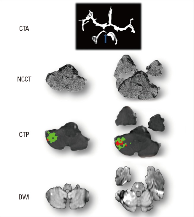
Figure 7.

Cerebellum Images of Case 7. Acute CTA shows, compared to normal left side, right superior cerebellar artery (SCA) is absent. No obvious change is observed on acute NCCT. CTP reveals hypoperfused lesion on SCA territory with the existence of penumbra (green area). After treatment, DWI shows stroke lesion (high signal) corresponding to infarct core of CTP (red area) with at least some of the penumbra saved.
