
Figure 9.
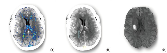
Cerebral images of Case 9. (A) acute DT map shows no specific perfusion lesion. (B) acute CTP source image shows hypodensity in the right lentiform (blue arrow). (C) follow-up DWI confirms the existence of ischemic lesion in the posterior right lentiform nucleus.