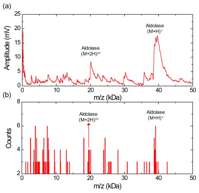
Figure 3.
(a) MALDI-TOF mass spectrum for aldolase with siniapinic acid matrix obtained using the silicon nanomembrane detector, showing singly and doubly charged aldolase ions, and a large number of fragment ions. (b) Histogram of the MALDI-TOF mass spectrum, showing a relative abundance of aldolase ions in the sample under test.

