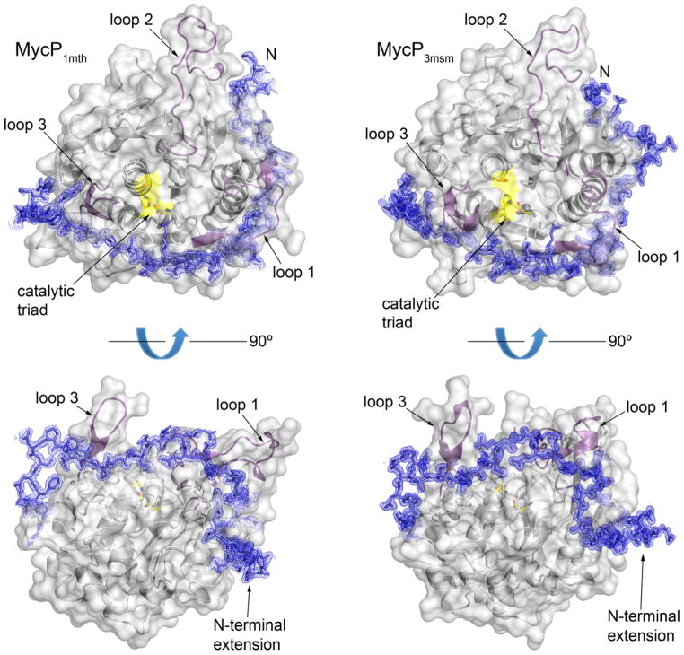
Fig. 4. The mycosin-specific N-terminal extension and loop insertions.
MycP1mth and MycP3msm structures are displayed as ribbons with semitransparent gray surface around the core subtilsin domain. The catalytic triad residues are highlighted in yellow. Loop insertions are highlighted in purple. The N-terminal extension amino acid residues are shown as blue sticks surrounded by σA-weighted 2FO–FC electron density map contoured at 1 σ.

