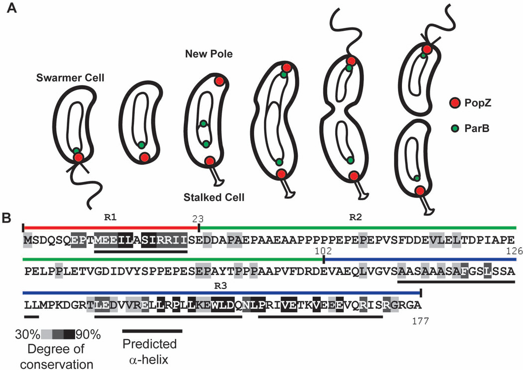
Figure 1.
Subcellular localization of PopZ as a function of the cell cycle and PopZ sequence.
A. A schematic of the Caulobacter cell cycle. Circles and theta structures within the cell indicate the replicating chromosome. Following the initiation of DNA replication, a new focus of PopZ assembles at the pole opposite the stalk, where it is required for tethering the newly duplicated ParB/parS centromere.
B. PopZ primary sequence, with shaded regions indicating the degree of conservation in Alphaproteobacteria. Black bars under the sequence represent predicted α-helices. The R1, R2, and R3 regions of the PopZ protein are indicated above the sequence as red, green, and blue bars, respectively.

