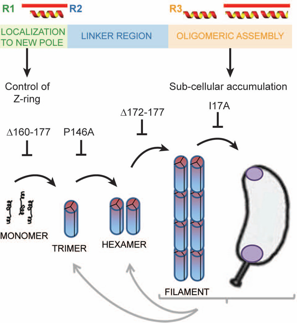
Fig. 8.
Functionally distinct regions of PopZ are responsible for control of cell division and the assembly of PopZ superstructures.
A. Schematic diagram of the PopZ protein sequence. Distinct functional regions, based on areas of conserved sequence, predicted positions of alpha helices, and our functional data, are color coded.
B. A model showing the relationship between PopZ assembly and sub-cellular localization. The first step is the self-association of monomers into rod-shaped trimers, which subsequently dimerize through lateral contact to form hexamers. End-to-end contacts between hexamers produce filaments, and in vivo, these filaments accumulate at cell poles. Each step in this process can be blocked by a mutation in PopZ, as indicated. Each of the forms is metastable and its frequency is influenced by protein concentration and conditions in the buffer or cell extract.

