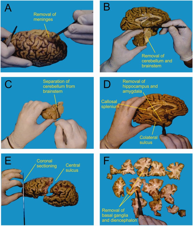
Figure 1.
After removing the meninges and blood vessels (A), the left brainstem and cerebellum were separated from the brain (B), and the cerebellar peduncles were cut to isolate the cerebellum (C). The hippocampal formation was removed out from the hemisphere by sectioning along the collateral sulcus until the caudalmost coronal level of the callosal splenium (D). The frontal lobe was then separated from the rest of the brain by a section made along the central sulcus (E), and cut coronally into slices of ∼1 cm thickness (E and F). Finally (F), the cerebral cortex was separated from the remaining regions (basal ganglia, diencephalon, mesencephalon, pons and medulla oblongata).

