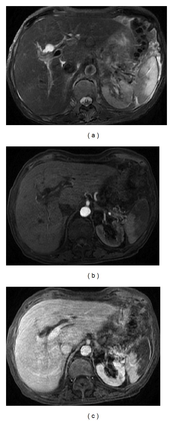
Figure 10.

Hematoma. Axial (a) T2wi SSFSE and postcontrast axial T1wi 3D-GRE with fat suppression in the arterial (b) and interstitial (c) phases. A chronic hematoma is depicted with a cystic appearance, regarded as a lesion with moderate hyperintensity on T2wi sequences (a) and hypointensity on T1wi with no perceptible enhancement ((b), (c)).
