
Figure 17.
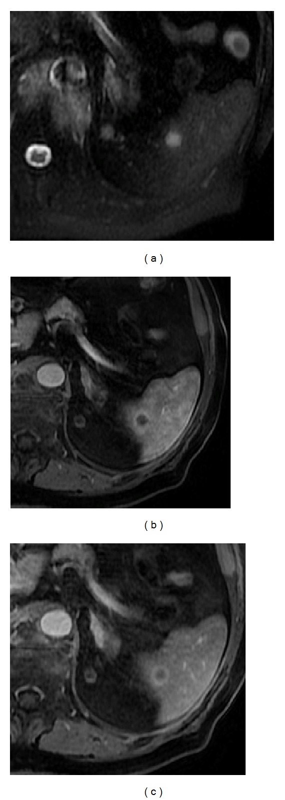
Splenic metastasis on a patient with a small-cell lung carcinoma. Axial T2wi SSFSE (a) and postcontrast axial 3D-GRE T1wi with fat suppression at the arterial (b) and venous (c) phases. Note the nodular lesion depicted as a hyperintense nodule on T2wi with peripheral ring-like enhancement.