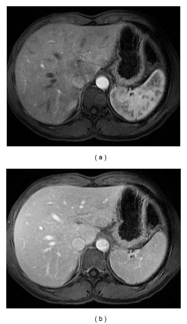
Figure 2.

Pattern of enhancement of the normal spleen. Postcontrast axial T1wi 3D-GRE with fat suppression in the arterial (a) and venous (b) phase. The spleen shows a heterogeneous pattern enhancement immediately after contrast material administration (a), secondary to differences in flow between the red and white pulps, becoming homogeneous in venous (b) and interstitial phases.
