
Figure 21.
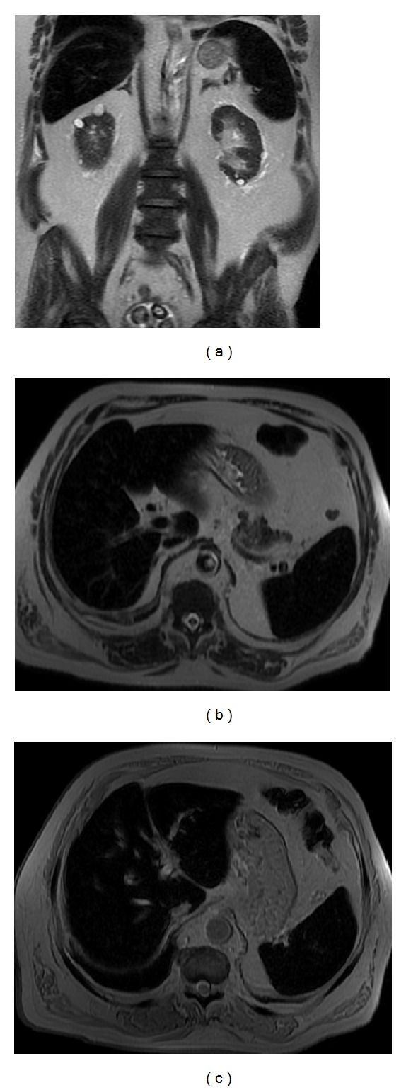
Paroxysmal nocturnal hemoglobinuria. Coronal (a) and axial (b) T2wi SSFSE and axial T2* (c) images. This patient with paroxysmal nocturnal hemoglobinuria shows diffuse diminished signal intensity of the liver and spleen on T2wi as a result of hemosiderin deposition. Notice the iron accumulation on the renal cortex (a).