Figure 4.
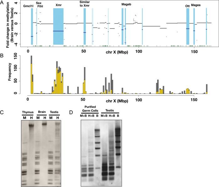
Demonstration of LoDs on the mouse X chromosome. (A) Differences in methylation levels between brain and testis genomic DNA along the mouse X chromosome. Fold changes in the methylation level, i.e. brain M-value versus testis M-value, were calculated for each CCGG segment, and plotted using the log2 scale along the X chromosome. The blue lines indicate genomic regions showing more than a 2-fold difference between the brain M-value and testis M-value. Light blue represents hypomethylated regions in the testis relative to the brain. Green dots at the bottom represent the positions of CGIs. (B) The positions of segmentally duplicated regions along the mouse X chromosome. Segmentally duplicated regions >1000 bases with >98% similarity are counted, and the frequencies of duplications (y-axis) are shown. (The data were from UCSC Genome Browser.) Grey bars represent duplications occurring on the X and other chromosomes, and yellow bars represent the frequencies of duplications mapped only on the X chromosome. (C) Methylation analysis of LoDs 10 and 12 by Southern blot hybridization. Genomic DNAs of the male thymus, male brain and testis were digested by either methylation-sensitive HpaII (H) or methylation-insensitive isoschizomer, MspI (M). The Southern blot was hybridized with a probe targeted to LoDs 10 and 12. A primer pair (FW: 5′-GCTGGGTCCAGCTTCCCTGG-3′, RV: 5′-TGGCACCCCTCCTGCCTGAT-3′) was used to amplify a 807-bp sequence using testis cDNA for generation of the probe. The 807-bp probe contains locally repeated sequences and corresponds to both LoDs 10 and 12 located upstream of Mageb1/b2 genes. (D) Methylation analysis of LoDs 10 and 12 in purified germ cells. Germ cells expressing the Mvh-Venus reporter were purified from adult testis by FACS. DNAs from purified germ cells and whole testis were digested with MspI plus BamHI (M + B), HpaII plus BamHI (H + B) or BamHI only (B). A Southern blot was made using these DNAs and hybridized with the same probe as used in Fig. 4C.

