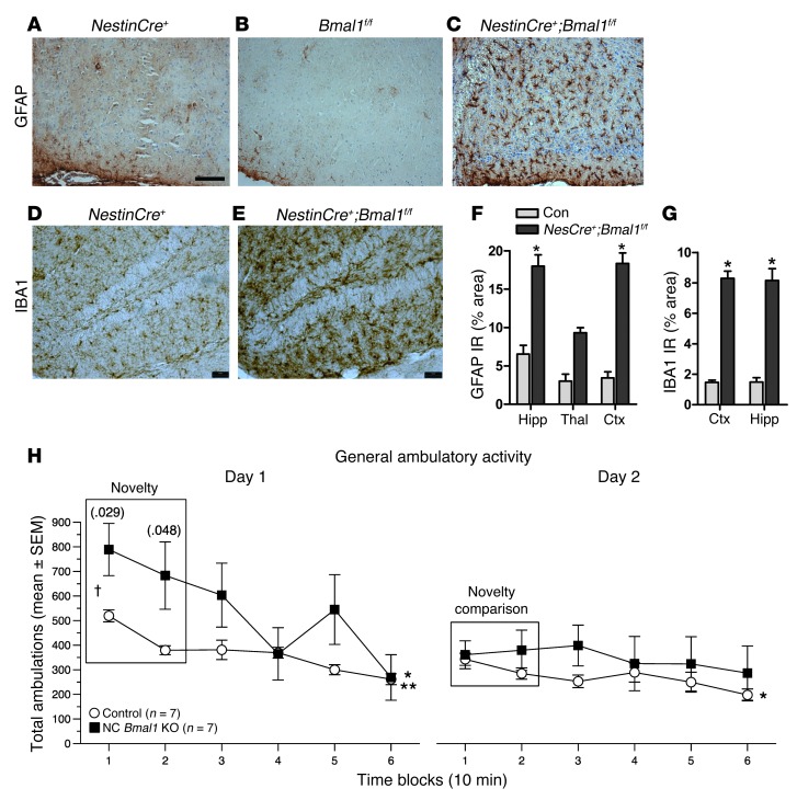
Figure 4. Brain-specific deletion of Bmal1 causes neuropathology and behavioral abnormalities.
GFAP staining shows marked astrocyte activation in the retrosplenial cortex of NestinCre+:Bmal1f/f mice (C), but not in NestinCre+ (A) or Bmal1f/f controls (B). Hippocampal microglial activation assessed by IBA1 immunoreactivity in a representative Cre+ control (D) and NestinCre+;Bmal1f/f mice (E). Scale bars: 200 μm. Quantification of GFAP (F) and IBA1 (G) immunoreactivity by percentage of area (n = 4 mice/genotype). *P < 0.05 versus control by 2-way ANOVA with Bonferroni’s post test. Ctx, cortex. (H) One-hour locomotor behavioral test reveals a significantly abnormal response to a novel environment in NestinCre+;Bmal1f/f mice (black squares) as compared with Bmal1f/f controls. Data for total ambulations are shown; similar data for vertical rearings are shown in Supplemental Figure 5. n = 7 mice/genotype. P values from repeated-measures ANOVA are displayed for novelty analysis. P < 0.05 for habituation analysis for both genotypes on day 1, but only for Bmal1f/f on day 2; *Bmal1f/f; **NestinCre+;Bmal1f/f.

