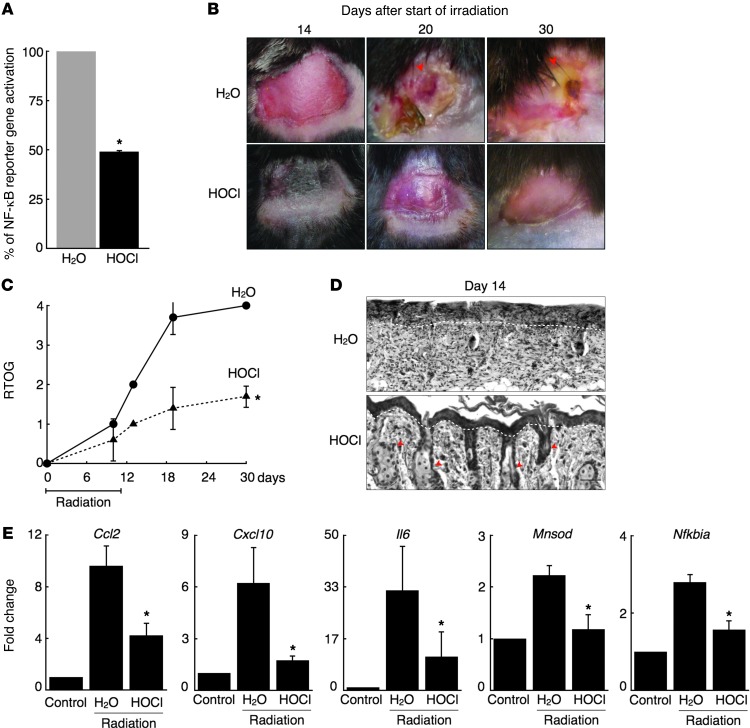
Figure 4. HOCl attenuates acute radiation dermatitis.
(A) Relative percentage of NF-κB reporter gene activation in the skin of LPS-stimulated NF-κB reporter mice treated topically with and without HOCl (n = 3 in each group). *P < 0.01. Four-week-old mice received 6 Gy irradiation daily for 10 days on their back skin. Prior to each dose of radiation, mice were treated with H2O or HOCl (n = 5–7 for each treatment group). (B) Images taken of the back skin on days 14, 20, and 30 after the start of irradiation. Representative images are selected from each group. (C) Score based on RTOG criteria for radiation-induced dermatitis in mice treated with H2O or HOCl. *P < 0.001 comparing treatment groups across 14-, 20,- and 30-day time points. P < 0.01 for individual 14-, 20-, and 30-day time points. (D) H&E staining of epidermis from indicated mice on day 14. White dashed line indicates basement membrane; red arrows indicate skin appendages. Scale bar: 20 μm. Representative images selected from each group. (E) Relative mRNA levels of Ccl2, Cxcl10, Il6, Mnsod, and Nfkbia in back skin isolated from irradiated mice treated with H2O or HOCl on day 14 (n = 4–6 for each treatment group). Control obtained from untreated mouse back skin. *P < 0.05 for all genes. Data are presented as the average ± SEM.

