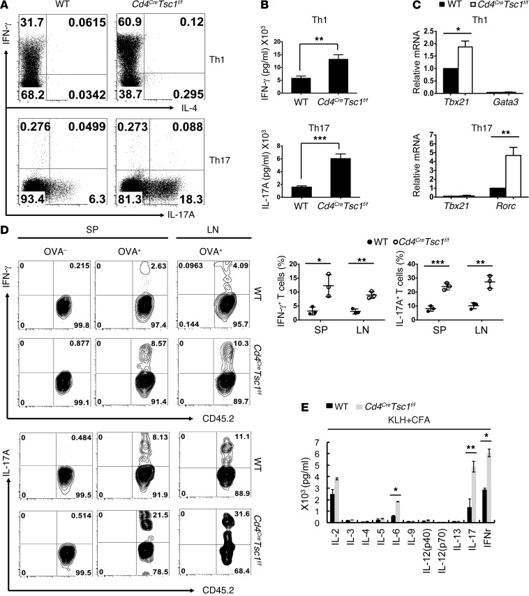
Figure 2. TSC1 deficiency promotes Th1 and Th17 differentiation.
(A) Naive CD4+ T cells from WT and Cd4CreTsc1f/f mice were cultured under Th1- and Th17-polarizing condition for 5 days. IFN-γ–, IL-4–, or IL-17A–producing cells were analyzed by intracellular cytokine staining (ICCS) 6 hours after restimulation with anti-CD3/CD28. (B) Th1 and Th17 cell cytokine production was measured by ELISA 24 hours after restimulation with anti-CD3/CD28. (C) RT-PCR analysis of mRNA expression levels of Th subset–specific transcription factors in polarized Th1 or Th17 cells. Data are representative of (A) or compiled from (B and C) three to five independent experiments. Error bars indicate the mean ± SD. *P < 0.05, **P < 0.01, and ***P < 0.001 by two-tailed, unpaired Student’s t test . (D) IFN-γ and IL-17 production (upper panel) and frequencies (lower panel) in T cells from recipient mice immunized with OVA/CFA. CD4+ T cells from WT or Cd4CreTsc1f/f OT-II mice were adoptively transferred into CD45.1 congenic B6 mice, followed by immunization with OVA/CFA. Splenocytes and lymph node cells obtained 6 days later were stimulated with OVA323–339 peptide for 24 hours and analyzed by ICCS and flow cytometry. (E) Immunization of WT and Cd4creTsc1f/f mice with KLH/CFA. CD4+T cells obtained 6 days later were stimulated with KLH for 72 hours, and the culture supernatants were analyzed by Bio-Plex multicytokine assay. Data are representative of (D) or compiled from (E) three independent experiments. Each symbol represents an individual mouse (n = 3). Error bars indicate the mean ± SD. *P < 0.05, **P < 0.01, and ***P < 0.001 by two-tailed, unpaired Student’s t test.

