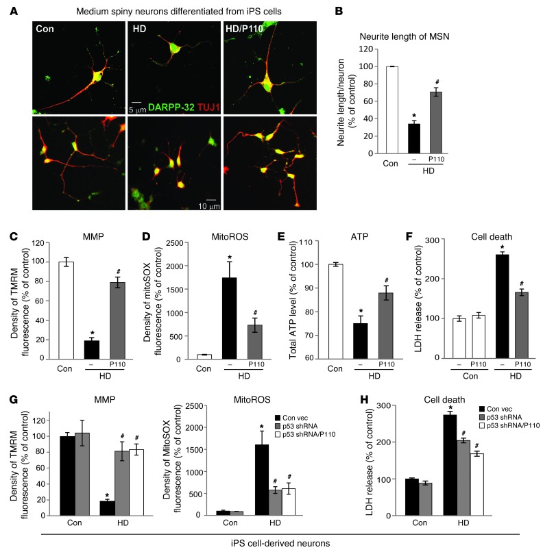
Figure 6. P110-TAT treatment reduced neurite shortening of MSNs from HD-iPS cells and corrected mitochondrial damage and cell death in patient neurons.
(A) Representative imaging on MSNs derived from Con- and HD-iPS cells. Top: A single neuron from each experimental group (original magnification, ×40). Bottom: A cluster of neurons at lower magnification (original magnification, ×20). (B) Data represent 3 independent experiments. At least 50 neurons/group were analyzed by an observed blinded to the experimental conditions. (C) Neuronal cells were labeled with TMRM fluorescence dye to indicate MMP. Density of TMRM red fluorescence only in cells with a neuronal-like morphology (multipolar cell bodies with at least 2 processes) was further quantitated as described in ref. 91. (D) Neuronal cells were stained with MitoSOX red, and density was analyzed in cells immunopositive for anti–DARPP-32. (E) Total ATP levels were measured using total lysates of mixed neuronal cells. Data represent 3 independent experiments. At least 50 neurons per group were analyzed. (F) After removal of BDNF for 48 hours, neuronal cell death was determined by the release of lactate dehydrogenase (LDH). Data represent 3 independent experiments. (G) Fluorescence density of EGFP-positive cells from neurons transduced by lentiviral particles containing p53 shRNA or control vector. Data represent 3 independent experiments. At least 50 EGFP-positive neurons per group were analyzed. (H) Neuronal cell death was determined by LDH release 2 days after removal of BNDF. Data are mean ± SEM. *P < 0.05 vs. Con; #P < 0.05 vs. HD.

