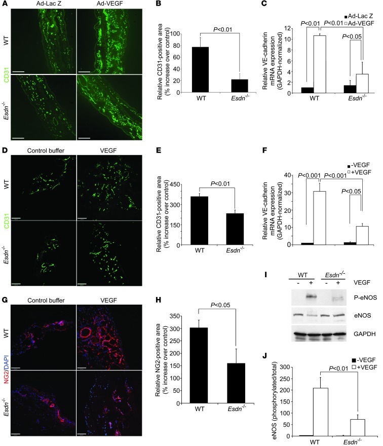
Figure 4. Attenuation of responses to exogenous VEGF in Esdn–/– mice.(A–C) Ear angiogenesis.
Examples (A) and quantification of CD31 immunostaining of ear blood vessels (B, n = 6) and ear GAPDH-normalized VE-cadherin mRNA expression (C, n = 3) 5 days following intradermal injection of Ad-VEGF or Ad-LacZ into opposite ears of WT and Esdn–/– mice. Scale bars: 100 μm. (D–H) Matrigel angiogenesis. Examples (D) and quantification of CD31 immunostaining (E, n = 6) and VE-cadherin mRNA (F, n = 3) of Matrigel plugs containing VEGF or control buffer implanted in WT and Esdn–/– mice. Scale bars: 100 μm. (G and H) Representative examples (G) and quantification (H) of NG2 pericyte immunostaining of Matrigels implanted in WT and Esdn–/– mice. Nuclei are stained with DAPI in blue. n = 3. Scale bar: 100 μm. (I and J) In vivo VEGF signaling. Example (I) and quantification (J, n = 3) of VEGF-induced lung eNOS phosphorylation in WT and Esdn–/– mice.

