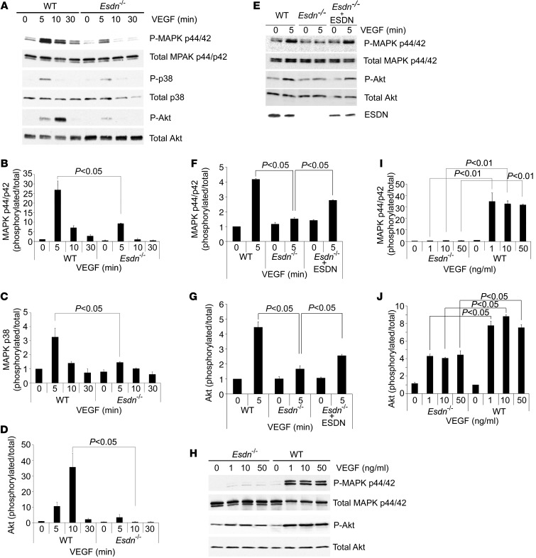
Figure 6. Attenuation of VEGF signaling in Esdn–/– MLECs.
(A–D) Western blot analysis of VEGF signaling in MLECs. Examples and quantification of VEGF-induced MAPK p44/42 Thr202/Tyr204 phosphorylation (A and B), p38 Thr180/Tyr182 phosphorylation (A and C), and Akt Ser473 phosphorylation (A and D) in WT and Esdn–/– MLECs. n = 3. (E–G) ESDN reconstitution in Esdn–/– MLECs. Example (E) and quantification (F and G) of Western blots of VEGF-induced MAPK p44/p42 Thr202/Tyr204 phosphorylation and Akt Ser473 phosphorylation following transient transfection of Esdn in Esdn–/– MLECs. (H–J) Western blot analysis of MAPK p44/42 Thr202/Tyr204 phosphorylation and Akt Ser473 phosphorylation in WT and Esdn–/– MLECs following stimulation with various concentrations of VEGF.

