
Figure 1.
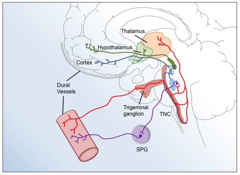
Representation of the trigeminovascular system in migraine. Trigeminal afferents innervate the dural vasculature. The first-order trigeminal neuron is located in the trigeminal ganglion. Its central terminal projects to the trigeminal nucleus caudalis (TNC) that extends from the dorsal medulla to the dorsal spinal horn of the first two cervical segments. Second-order neurons of the TNC project to the posterior thalamus. The sphenopalatine ganglion (SPG) also provides reflex parasympathetic innervation to dural vessels. This illustration has been adapted from (Goadsby et al., 2002).