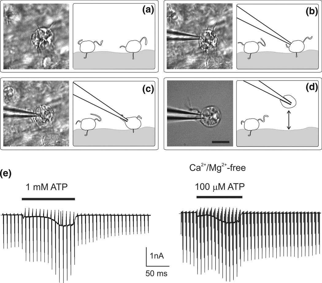
FIGURE 3.
ATP triggers P2X7receptor-mediated currents in amoeboid microglial cells collected from the surface of corpus callosum slices of young (P5–P7) mice. (a–d) Method of patch-voltage clamping of single amoeboid microglial cells from the surface of corpus callosum slice. Combinations of images taken by means of infrared video microscopy (left panels) and schematic drawings showing the method of cell patch-clamping. Video images were captured with an intensified CCD camera (Hamamatsu, Japan) and digitized by a frame grabber connected to the PC. (a) Initial position of amoeboid microglial cells situated on the surface of a corpus callosum slice. (b) A single microglial cell was approached with a micropipette and a whole-cell patch clamp configuration was established. (c) After 2–3 min, the cell partially spread over the pipette, intensifying the cell-to-pipette contact. (d) Finally, the cell was lifted for 200 µm over the slice surface. Scale bar in (d) = 10 µm. (e) P2X7-mediated membrane currents measured from amoeboid microglial cells in control conditions and Ca2+/Mg2+-free bath solution. (Reprinted with permission from Ref 79. Copyright 1996 Elsevier)

