
Figure 1.
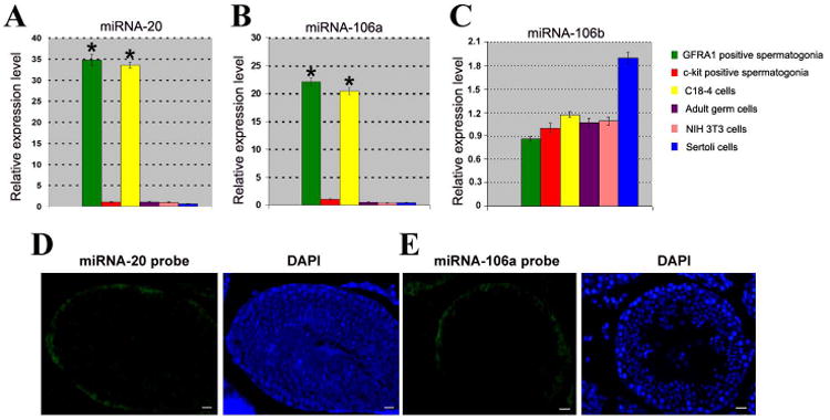
Distinct expression of miRNAs in various types of cells and subcellular localization in adult mouse testes. (A-C) Real time PCR showed the expression of miRNA-20, miRNA-106a, and miRNA-106b in GFRα1 positive spermatogonia, c-kit positive spermatogonia, C18-4 cells, adult male germ cells, NIH 3T3 cells, and Sertoli cells. House keeping miRNA-16 served as a loading control of total miRNAs. To compare the expression of miRNA-20, miRNA-106a and miRNA-106b in different types of cells, the expression of these miRNAs in c-kit positive spermatogonia was set as 1. Compared to c-kit positive spermatogonia, “*” indicated significant difference (P<0.05). (D-E) Fluorescein in situ hybridization displayed restricted expression of miRNA-20 (D) and miRNA-106a (E) only to spermatogonia at the basement membrane of the seminiferous tubules in adult mouse testes. DAPI was used to show cell nuclei. Scale bars = 20 μm (D, E).