
Figure 1. This figure shows example axial slices from a typical subject scanned at birth (left column), 2 (middle column), and 4 years old (right column).
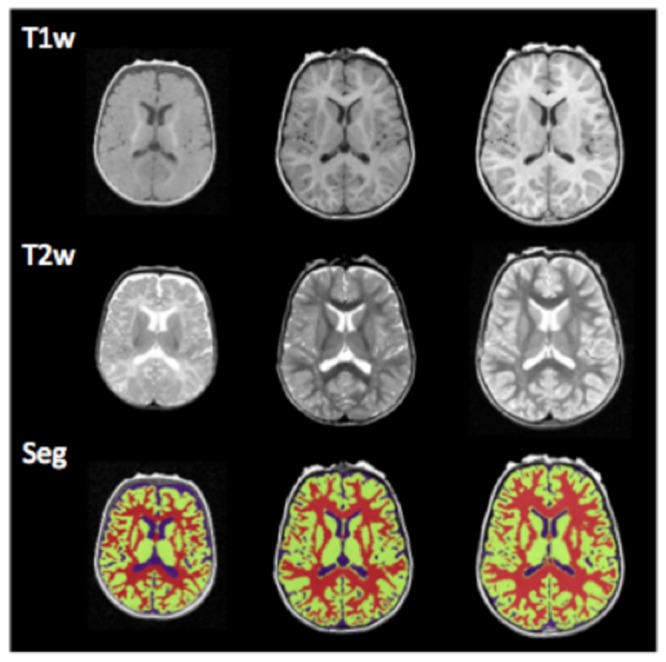
T1 MRI, T2 MRI, and segmented gray matter (green) and white matter (red) are provided.
Official websites use .gov
A
.gov website belongs to an official
government organization in the United States.
Secure .gov websites use HTTPS
A lock (
) or https:// means you've safely
connected to the .gov website. Share sensitive
information only on official, secure websites.
T1 MRI, T2 MRI, and segmented gray matter (green) and white matter (red) are provided.