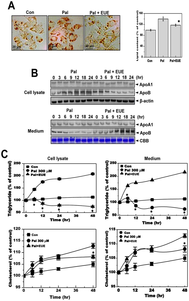
Figure 2. E. ulmoides Oliver extract reduces hepatic lipid accumulation and levels of secreted apolipoprotein B.
(A) Cells were treated with 300 μM palmitate in the absence or presence of 100 μg/mL EUE for 12 hours. Fat accumulation was determined by Oil Red O staining. Images of cells were obtained at 200X original magnification and used for quantitative analysis of cellular lipid deposition (right). * p<0.05, significantly different from cells treated with palmitate alone. (B) Cells were treated with 300 μM palmitate in the presence or absence of 100 μg/mL EUE for 0, 3, 6, 9, 12, 18, or 24 hours. Cell lysates and media samples were subjected to immunoblot analysis with anti-ApoA1 or anti-ApoB. CBB staining was performed as an equal loading control. (C) Cells were treated with 300 μM palmitate in the absence or presence of 100 μg/mL EUE for 0, 6, 12, 24, or 48 hours. Triglyceride and cholesterol levels were measured for both cell lysates and media alone. * p<0.05, significantly different from cells treated with palmitate alone at the corresponding time point. Pal, palmitate; EUE, E. ulmoides Oliver extracts; CBB, Coomassie brilliant blue.

