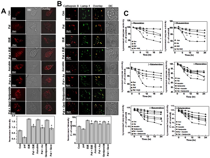
Figure 5. E. ulmoides Oliver extract, aucubin, and geniposide enhance lysosomal activity.
(A) Cells were treated with 100 μg/mL EUE, 10 μg/mL aucubin, or 10 µg/mL geniposide in the presence or absence of 300 µM palmitate for 12 hours, followed by exposure to 5 μM LysoTracker and image acquisition and quantification (lower panel). (B) Cells were fixed and immunostained with antibodies for cathepsin B or Lamp-1. Quantification of fluorescence is shown (lower). (C) The activities of α-mannosidase, β-galactosidase, and β-glucuronidase were analyzed from lysosomal extracts of cells treated for the indicated time periods. * p<0.05, significantly different from cells treated with palmitate alone (A, B); * p<0.05, significantly different from cells treated with palmitate alone at each corresponding time point (C). DIC, differential interference contrast microscopy; Pal, palmitate; EUE, E. ulmoides Oliver extract.

