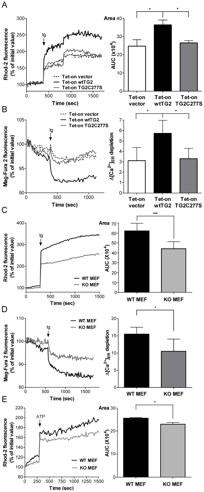
Figure 3. Transglutaminase 2 enhances both the Ca2+ release from the endoplasmic reticulum and the mitochondrial Ca2+ uptake.
Tet-on vector, Tet-on wtTG2 and Tet-on TG2C277S cells treated with Dox (50 µM) for 18 h were exposed to 5 µM thapsigargin (tg), and changes in the Ca2+ concentrations in the mitochondria (A) or in the ER (B) were monitored as described in the Materials and Methods. Wild type and TG2KO MEF cells were exposed to 5 µM thapsigarginin Ca2+ free medium and changes in the Ca2+ concentrations in the mitochondria (C) or in the ER (D) were monitored as described in the Materials and Methods. (E) Wild type and TG2 KO MEF cells were exposed to 500 µM ATP in Ca2+-free medium, and changes in the intra-mitochondrial Ca2+ concentrations were monitored as described in the Materials and Methods. Left panels, Representative kinetic average changes in mitochondrial or ER Ca2+signals induced by thapsigarginor ATP over time are shown. Right panels, Areas, statistical evaluation of integrated Ca2+ responses are shown. AUC, area under the curve. These data are representative of at least three experiments and shown as mean ± SD. *P<0.05; ***, P<0.001.

