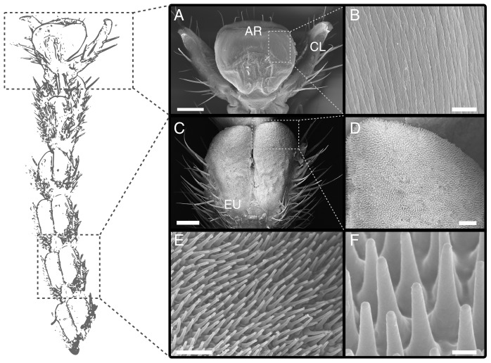
Figure 3. Morphology of the tarsus of Carausius morosus.
(A) pre-tarsus with adhesive pad (arolium) between the pair of claws. (B) surface of the arolium contact area with folds running along the proximal-distal axis. (C) pair of euplantulae on the ventral side of the tarsus (second tarsal segment). (D)–(F) acanthae on the surface of the euplantulae. AR arolium, CL claw, EU euplantulae. Scale bars are (A) 200  , (B) 4
  (C) 100 m, (D) 20
  , (E) 1
  , (F) 10
  .
.

