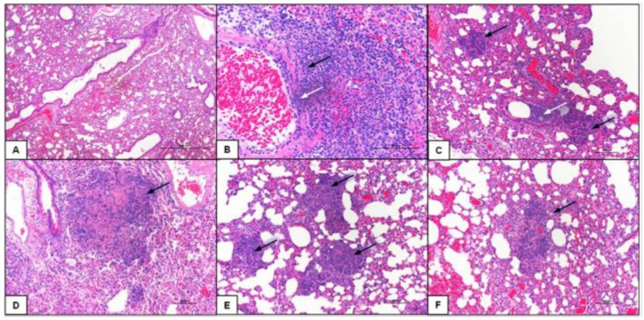
Supplemental Figure 1.
Representitve examples of lung tissue pathology seen in infected BALB/c mice. Representative images of Hematoxylin and Eosin stained mouse lungs sections that highlight the following pathology: normal lung (A); Perivasculitis (black arrow) and vasculitis with neutrophil component (white arrow) (B); scattered microabscesses (black arrows) and cell debris in bronchial lumen with neutrophil component (white arrow) (C); areas of necrosis (black arrows) (D); microabscesses (black arrows) (E); and small necrotic foci (black arrow) (F); Magnification 40× and scale bar = 100µM.

