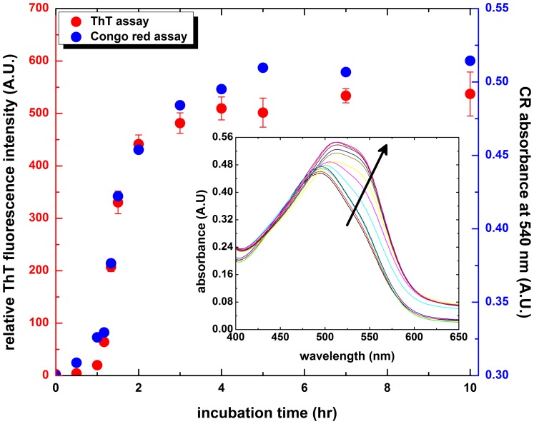
Figure 1. Amyloid fibrillogenesis kinetics of hen egg-white lysozyme (HEWL).
The extent of fibril formation was monitored via Congo red absorbance at 540(ThT) fluorescence as a function of incubation time. HEWL samples were dissolved in 100 mM glycine buffer (pH 2.0) and incubated at 55°C with agitation of 580 rpm during the course of experiment. The extent of fibril formation was also monitored via Congo red absorption spectra shown in the inset (the arrow indicates the increasing incubation time). Data represent the mean ThT fluorescence intensity of at least 5 independent experiments (n≥5). Error bars represent the standard deviation (S.D.) of the fluorescence measurements.

