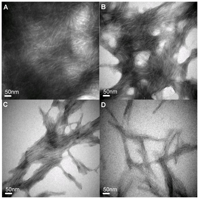
Figure 3. Transmission electron micrographs of HEWL samples with various concentrations of carnosine.
Negatively stained electron micrographs of (A) HEWL alone, (B) HEWL co-incubated with 20 mM carnosine, (C) HEWL co-incubated with 40 mM carnosine, and (D) HEWL co-incubated with 50 mM carnosine. HEWL samples in the absence and presence of carnosine were prepared at pH 2.0 and incubated for 10 hr. The scale bar represents 50 nm.

