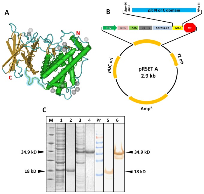
Figure 1. Construction of the recombinant expression vector pRSET A-PLC_N/C and cloning and expression of recombinant N and C-domains.
(A) Crystal structure of C. perfringens phospholipase C protein (PDB ID 1MOX) drawn by Cn3D software. The ten amino acid linker GNDPSVGKNV region is highlighted in blue. (B) Schematic diagram of plasmid construct. Gene sequences corresponding to plc N or C-domain were inserted into pRSET A for the expression of recombinant protein. (C) Coomassie Blue stained 12% SDS-PAGE and western blot analysis of whole cell lysates of IPTG induced N and C-domain transformant E. coli cells. Arrow mark indicates the expressed recombinant proteins. Blot was probed with commercial anti-PLC polyclonal antibodies. M-Unstained protein marker; 1-induced recombinant C clone; 2- purified C-domain; 3-induced recombinant N clone; 4-purified N domain; PrPr-prestained protein ladder; 5-C-domain; 6-N-domain.

