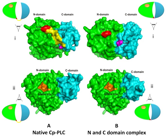
Figure 4. Surface representation of structures of native alpha toxin from C. perfringens and N and C-domain hybrid.
A. Crystal structure of native alpha toxin from C. perfringens illustrating N and C-domains along with a linker (View i) and the active site (View ii). B. Docking study of N and C-domains presenting configuration changes during interaction (View i) and the unchanged active site on N domain (View ii). The N-domain and C-domain are colored in green and cyan respectively. The last residue of N and first residue of C are displayed in red and pink colors, respectively. The residues of linker region and active site are presented in yellow and orange colors, respectively.

