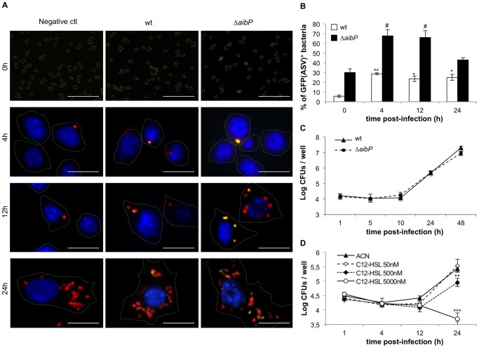
Figure 4. B. melitensis both produces and degrades long-chain AHLs during macrophage infection.
(A and B) B. melitensis wt and ▵aibP QS reporter strains were used to infect monolayers of RAW264.7 murine macrophages. Prior to (0h) and during infection, bacteria or cells were fixed, bacteria were labelled with a monoclonal A76-12G12 anti-LPS antibody and DNA was labelled with DAPI. (A) Immunofluorescence micrographs are representative from at least two independent experiments. Bacteria LPS appears in red, DNA in blue. Scale bar, 10 µm. (B) The percentage of GFP(ASV)-positive bacteria at different times post-infection was determined as described in the Material and Methods section. Error bars represent the standard deviation from two independent experiments. Data have been analyzed by ANOVA I after testing the homogeneity of variance (Bartlett). * and ** denote significant differences (P < 0.05 and P < 0.01) in relation to wt bacteria prior to infection (0h) while # denotes a significant difference (P < 0.05) in relation to ▵aibP bacteria prior to infection. (C) Intracellular replication of B. melitensis wt and ▵aibP strains in RAW264.7 murine macrophages. At indicated times, cells were lysed and intracellular colony forming units (CFUs) were determined. Error bars represent the standard deviation of triplicates in one representative experiment out of three. (D) RAW264.7 macrophages were infected with B. melitensis wt in the presence of C12-HSL or ACN (negative control) and treated as described (C). ** and *** denote significant (P < 0.01 and P < 0.001 respectively) differences in relation to infection by wt bacteria in the presence of CAN (Bartlett and ANOVA I analysis).

