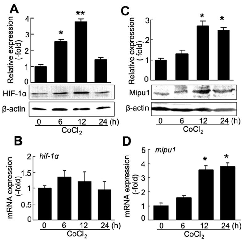
Figure 1. CoCl2 induces HIF-1α and Mipu1 expression in H9C2 cells.
A: immunoblotting detection for CoCl2-induced expression of HIF-1α protein (n=3), densitometry analysis of HIF-1α band against β-actin band sown in upper panel. * p<0.05, ** p<0.01 versus “0” (CoCl2 untreated cells). B: Quantitative real time PCR detection for the expression of hif-1α induced by CoCl2 (n=4. triplicate for each sample). C: immunoblotting detection for CoCl2-induced expression of Mipu1 protein (n=3), densitometry analysis of Mipu1 band against β-actin band shown in upper panel. * p<0.05 versus “0” (CoCl2 untreated cells). D: Quantitative real time PCR detection for the expression of mipu1 induced by CoCl2 (n=4. triplicate for each sample), compared with CoCl2 untreated cells, * p<0.05.

