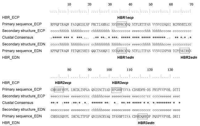
Figure 1. Sequence alignment of human eosinophil RNases and HBRs.
Amino acid sequences of human ECP (accession number P10153.2) and EDN (accession number P12724.2) were aligned using Clustal X2 [54]. Putative heparin binding regions (HBRs) were framed. Fully conserved amino acids were indicated by asterisk (*), highly similar amino acids were indicated by colon (:), and weakly similar amino acids were indicated by dot (.). Secondary structures of human eosinophil RNases were shown in the middle. Structural elements including α-helices, β-strands, and coils were represented as “h”, “e”, and “c”, respectively.

