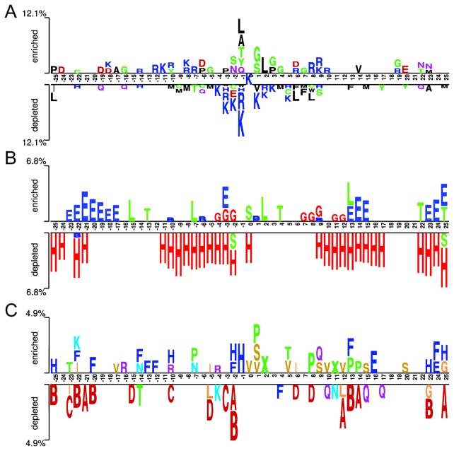
Figure 3. The two-sample logo illustration of the context (sequence neighbors) of ubiquitination sites.
(A) The positional residue pattern; (B) the secondary structure pattern and (C) the local conformation (structural alphabet) pattern where a seven-group color palette is used: helix (red), helix-like (orange), strand (blue), highly curved coil (yellow), moderately curved coil (violet) and flat coil (green). See also Tables 1 and 2 for the description of the secondary structure type and structural alphabet state, respectively.

