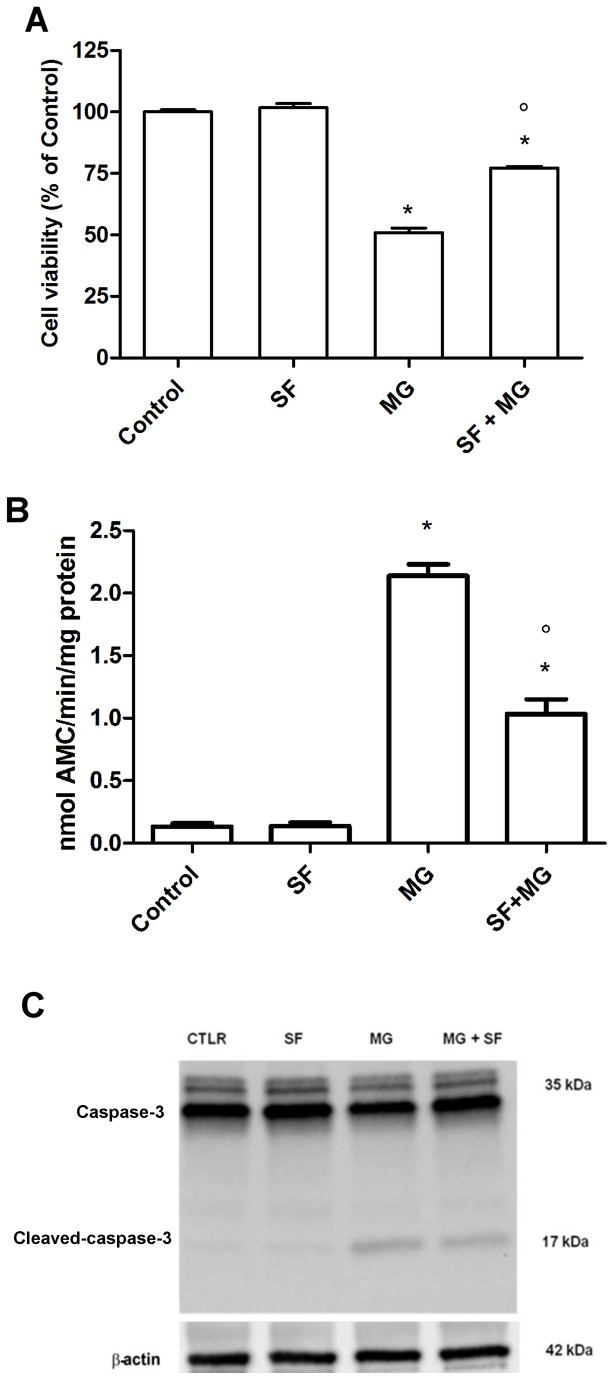
Figure 5. SF protection against MG-induced damage.
Cardiomyocytes were treated with 5 µM SF for 24 h before addition of 1 mM MG. (A) Cell viability was assessed by MTT assay and reported as percent cell viability in comparison to control. (B) Caspase-3 activity was measured spectrofluorimetrically by hydrolysis of the peptide substrate acetyl-Asp-Glu-Val-Asp-7-amido-4-methylcoumarin (Ac-DEVD-AMC). (C) Cell lysates were immunoblotted with caspase-3 antibody that detects both full length caspase-3 (35 kDa) and the large fragment of caspase-3 resulting from cleavage (17 kDa). Data are presented as mean ± SD, n = 4 in each group,* p<0.05 vs Control; ° p<0.05 vs MG.

