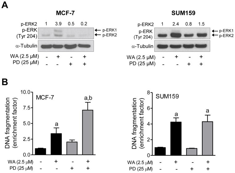
Figure 4.
The effect of pharmacological inhibition of ERK on WA-mediated apoptosis in human breast cancer cells. The MCF-7 and SUM159 human breast cancer cells were pretreated with 25 μM PD98059 for 1 h, and then exposed to 2.5 μM WA in the absence or presence of PD98059 for an additional 24 h. (A) Western blotting for phosphorylated ERK. Quantitation relative to DMSO-treated cells is shown. (B) Histone-associated DNA fragment release into the cytosol. Quantitation relative to DMSO-treated cells is shown. Combined results (n = 6) from two independent experiments are shown as mean ± S.D. Statistical significance was determined by one-way ANOVA with Bonferroni’s multiple comparison test. aSignificantly different (P<0.05) compared with respective control. bSignificantly different (P<0.05) between groups at the same dose.

