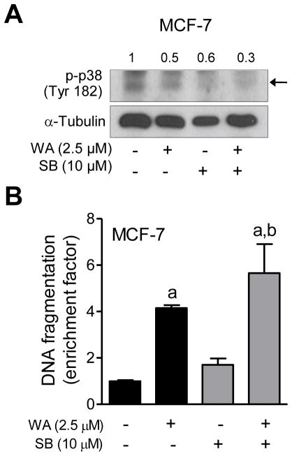
Figure 6.
The effect of pharmacological inhibition of p38 MAPK on WA-mediated apoptosis in MCF-7 cells. The MCF-7 cells were pretreated with 10 μM SB202190 for 1 h, exposed to 2.5 μM WA in the absence or presence of SB202190 for 24 h, and then processed for western blot analysis or apoptosis detection. (A) Western blotting for phosphorylated p38 MAPK and cleaved PARP. Quantitation for phosphorylated p38MAPK relative to DMSO-treated cells is shown. (B) Histone-associated DNA fragment release into the cytosol. Quantitation relative to DMSO-treated cells is shown. Combined results (n = 6) from two independent experiments are shown as mean ± S.D. Statistical significance was determined by one-way ANOVA with Bonferroni’s multiple comparison test. aSignificantly different (P<0.05) compared with respective control. bSignificantly different (P<0.05) between groups at the same dose.

