
Fig. 4.
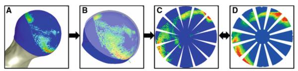
Process for comparing experimental and FE results pixel-wise. a FE results were extracted from the articular surface of the femoral head. b FE results were projected to a sphere, and the experimental position of the pressure-sensitive film is overlaid. c The spherical projection was mapped to a planar rosette, creating a simulated rosette with the same dimensions as the experimental rosette. d The experimental rosette was used as the reference standard for pixel-wise comparison against the simulated rosette