Abstract
Long-term nicotine exposure induces alterations in dopamine transmission in nucleus accumbens (NAcc) that sustain the reinforcing effects of smoking. One approach to understand the adaptive changes that arise involves measurement of endogenous dopamine release using voltammetry. We therefore treated rats for 2-3 months with nicotine and examined alterations in nAChR subtype expression and electrically-evoked dopamine release in rat NAcc shell, a region key in addiction. Long-term nicotine treatment selectively decreased stimulated α6β2* nAChR-mediated dopamine release compared to vehicle-treated rats. It also reduced α6β2* nAChRs, suggesting the receptor decline may contribute to the functional loss. This decreased response in release after chronic nicotine treatment was still partially sensitive to the agonist nicotine. Studies with an acetylcholinesterase inhibitor demonstrated that the response was also sensitive to increased endogenous acetylcholine. However, unlike the agonists, nAChR antagonists decreased dopamine release only in vehicle- but not nicotine-treated rats. Since antagonists function by blocking the action of acetylcholine, their ineffectiveness suggests that reduced acetylcholine levels partly underlie the dampened α6β2* nAChR-mediated function in nicotine-treated rats. Since long-term nicotine modifies dopamine release by decreasing α6β2* nAChRs and their function, these data suggest that interventions that target this subtype may be useful for treating nicotine dependence.
Keywords: addiction, nicotine, nicotinic receptors, nucleus accumbens, voltammetry
Nicotine addiction is the most common form of chemical dependence in the United States, with ~25% of the adult population using tobacco products despite their well known adverse health effects (Benowitz 2010). Although many smokers would like to quit, the vast majority are back to smoking within a week of cessation (Zhu et al. 2012). These are disturbing statistics in view of the significant health and economic burdens of smoking. Several smoking cessation medications exist; however, the best one-year abstinence rates are still only ~25% (Dhelaria et al. 2012, Mills et al. 2012, Zhu et al. 2012, Keating & Lyseng-Williamson 2010). Identification of the nAChRs and neurobiological processes that maintain nicotine dependence are thus important as such work will help in the development of more successful smoking cessation strategies.
It is well known that nicotine is the main addictive component of tobacco products. Like other drugs of abuse, nicotine enhances dopamine transmission in the mesolimbic dopamine pathway, which is thought to play a critical role in the reinforcing effects that initiate and maintain nicotine dependence (Tuesta et al. 2011, Corrigall et al. 1992, Balfour 2009, De Biasi & Dani 2011, Di Chiara et al. 2004). Extensive studies using genetically modified mice show that nicotine's effects are mediated through an interaction at neuronal nicotinic receptors (nAChRs) of which there are multiple subtypes (Changeux 2010). Converging evidence suggests that both α4β2* and α6β2* nAChRs in the mesolimbic dopaminergic pathway mediate numerous aspects related to nicotine reward, as well as withdrawal (Brunzell 2012, Epping-Jordan et al. 1999, Gotti et al. 2010, Jackson et al. 2009, Picciotto et al. 1998, Pons et al. 2008, Tapper et al. 2004). It was initially somewhat difficult to reconcile these research findings with voltammetry data which suggested that α6β2* nAChRs are the prominent regulators of dopamine release in NAcc (Exley et al. 2008, Perez et al. 2012). However, recent reports suggest that the α6α4β2* nAChR population may be the one important for nicotine's effects on dopamine transmission. Support for this idea comes from studies showing that α6α4β2* nAChRs primarily modulate dopamine neuron firing in the ventral tegmental area (VTA) and endogenous dopamine release in the NAcc (Exley et al. 2011, Liu et al. 2012, Zhao-Shea et al. 2011). In addition, long-term nicotine exposure decreases α6α4β2* nAChR expression (Perez et al. 2008a) providing further evidence for a role of this receptor subtype in nicotine dependence.
The purpose of this study was to further understand how long-term nicotine treatment modifies mesolimbic dopaminergic function by examining adaptive changes in dopamine release using cyclic voltammetry. We focused on the accumbens shell since dopamine release in this region has been linked to the rewarding or reinforcing properties of nicotine while release in the accumbens core and striatum is more highly associated to the presentation of a conditioned stimulus (Balfour 2009, Changeux 2010, Sellings et al. 2008, Wise 2009). We also measured nAChR subtype levels using receptor autoradiography. The results show that long-term nicotine exposure modulates electrically-evoked dopamine release in the NAcc by reducing α6β2* nAChR levels and their function. These data suggest that drugs that stimulate α6β2* nAChRs may be of benefit in treating nicotine addiction.
Materials and methods
Animal treatment
Adult male Sprague-Dawley rats (220-250 g) purchased from Charles River Laboratories (Gilroy, CA) were placed in a temperature-controlled room with a 12 h dark/light cycle and housed 3-4 per cage. All animals had free access to food and water. After several days of acclimation, rats were given drinking water containing 1% saccharin (Sigma Chemical Co., St. Louis, MO), which was used as a vehicle to mask the bitter taste of nicotine. They were then randomly divided into two treatment groups 3 d later. One group was maintained on saccharin only. Nicotine was added to the saccharin-containing solution of the other group at a concentration of 25 μg/ml nicotine (free base). Nicotine treatment was given for 2-3 months, as other studies have shown that the changes that arise during such a time period may model those in long-term smokers (Zhang et al. 2011). Weights were monitored twice a week, with no significant differences between the two treatment groups. The rats were killed by decapitation using a guillotine. All procedures conform to the NIH Guide for the Care and Use of Laboratory Animals and were approved by the Animal Care and Use Committee of SRI International.
Plasma Cotinine levels
Blood was drawn from the lateral saphenous vein under isofluorane anesthesia 2-3 wk after the rats were on nicotine treatment. Plasma cotinine was determined using an EIA kit (Orasure Technologies, Bethlehem, PA). Cotinine is a long-lasting nicotine metabolite used as an index of nicotine intake. The plasma cotinine levels in nicotine-treated rats were 358 ± 29 ng/ml (n=10), which are comparable to those in the plasma of smokers (Matta et al. 2007). For some animals, plasma was also collected after 2 months of treatment, with cotinine values similar to samples from the first blood draw. No rats were excluded due to low cotinine levels. There was no detectable plasma cotinine in saccharin-treated rats.
Tissue preparation
The brain was quickly removed and chilled in ice-cold, pre-oxygenated (95% O2/5% CO2) buffer containing: 30 mM NaCl, 4.5 mM KCl, 1.2 mM NaH2PO4, 1.0 mM MgCl2, 10 mM glucose, 26 mM NaHCO3, and 18 mM sucrose (pH 7.4). Coronal slices containing the NAcc (350 μm thick) were cut using a vibratome (Leica VT1000S) in the same buffer. Slices were then incubated for at least 1 hour in oxygenated physiological buffer containing: 125 mM NaCl, 2.5 mM KCl, 1.2 mM NaH2PO4, 2.4 mM CaCl2, 1.2 mM MgCl2, 10 mM glucose, and 26 mM NaHCO3 (pH 7.4) at room temperature. Each slice was transferred to a submersion-recording chamber (Campden Instruments Ltd., Lafayette, IN), perfused at 1 ml/min with 30° C, oxygenated buffer, and allowed to equilibrate for 30 min.
Some brains were immediately quick frozen in isopentane on dry ice and stored at –80o C. Sections (8 μm) were prepared using a cryostat (Leica Microsystems, Inc., Deerfield, IL) cooled to –20o C. Frozen sections were thaw-mounted onto Superfrost Plus slides (Fisher, Pittsburgh, PA), air-dried and stored at −80° C for nAChR autoradiography.
Cyclic Voltammetry
Carbon fiber microelectrodes (7 μm in diameter; tip length ~100 μm) were constructed as previously described (Perez et al. 2008b). The electrode was positioned below the surface of the slice and its potential linearly scanned every 100 ms from 0 to −400 to 1000 to −400 to 0 mV versus an Ag/AgCl reference electrode at a scan rate of 300 mV/ms. Only the carbon fiber was inserted into the slice to avoid tissue damage by the glass. Current was recorded and digitized at a frequency of 50 kHz with an Axopatch 200B amplifier (Molecular Devices, Sunnyvale, CA). Triangular wave generation and data acquisition were controlled by pClamp 9.0 software (Molecular Devices, Sunnyvale, CA). Background current was digitally subtracted to obtain the voltammograms used for the identification of dopamine (confirmed by an oxidation peak ~500-600 mV and a reduction peak around −200 mV). Peak oxidation currents were converted into concentrations after post-experimental calibration of the electrode with fresh solutions of 0.5-1.0 μM dopamine. Dopamine release was measured as the maximal peak response obtained after electrical stimulation.
Electrically-evoked dopamine release was measured in the dorsal half of NAcc shell. Electrical stimulation was applied using a bipolar stimulating electrode (Plastics One, Roanoke, VA) connected to a linear stimulus isolator (WPI, Saratoga, Fl) and triggered by a Master-8 pulse generator (A.M.P.I., Jerusalem, Israel). The stimulating electrode was consistently placed so that it just touched the surface of the slice and the carbon fiber electrode was positioned ~100 μm away. Evoked release was elicited by either a single electrical pulse or a train of 4 pulses (1-4 ms in duration) at 30 Hz applied every 2.5 min. This stimulation paradigm was based on our previous studies showing similar drug effects with a 30 Hz and a 100 Hz stimulation frequency (Perez et al. 2010). In addition, previous reports show that burst firing of rat dopamine neurons in vivo occurs at approximately 20 Hz (Zhang et al. 2009a). Control evoked release was assessed in physiological buffer. NAChR-modulated release was assessed in the presence of 100 nM α-conotoxinMII (α-CtxMII) to antagonize α6β2* nAChRs followed by 100 μM mecamylamine (Mec) or 100 nM dihydro-β-erythroidine (DHβE) to block all nAChR subtypes. The effects of Mec or DHβE alone are similar to that of either antagonist plus α-CtxMII (data not shown). Superfusion of the slice with α-CtxMII maximally decreased release within ~15 min and responses were recorded over a 1 h period. Responses in the presence of Mec and DHβE were recorded over at least a 2 h period. Slices were also exposed to nicotine (300 nM) and signals recorded over at least a 1 h period to determine how release was altered under this treatment condition. To determine the effects of acetylcholinesterase inhibition, slices were superfused with ambenonium dichloride at increasing concentrations (0.1, 1, 10, 40, 80, 100 and 1000 nM). All drug concentration used have been shown to yield a maximal effect (Exley et al. 2008, Zhou et al. 2001). The changes in release observed with all drugs were reversible upon prolonged wash (1-2 h). The reported effects represent the average of those signals obtained once a stable maximal response was established.
125I-Epibatidine Autoradiography
Binding of 125I-epibatidine (2200 Ci/mmol; Perkin Elmer Life Sciences, Boston, MA, USA) was done as previously reported (Quik et al. 2000). Slides were pre-incubated at 22° C for 15 min in buffer containing 50 mM Tris, pH 7.5, 120 mM NaCl, 5 mM KCl, 2.5 mM CaCl2, and 1.0 mM MgCl2. They were incubated for 40 min with 0.015 nM 125I-epibatidine in the presence or absence of α-CtxMII (100 nM). They were then washed, dried and exposed to Kodak MR film with 125I-microscale standards (GE Healthcare, Chalfont St. Giles, Buckinghamshire, UK) for several days. Nonspecific binding was assessed in the presence of 100 μM nicotine and was similar to the film blank.
125I-α-CtxMII Autoradiography
Binding of 125I-α-CtxMII (specific activity, 2200 Ci/mmol) was done as reported previously (Quik et al. 2001). Striatal sections were preincubated at room temperature for 15 min in binding buffer (144 mM NaCl, 1.5 mM KCl, 2mM CaCl2 1mM MgSO4, 20mM HEPES and 0.1% bovine serum albumin, pH 7.5) plus 1 mM PMSF (phenylmethylsulfonyl fluoride). This was followed by 1 h incubation at room temperature in binding buffer containing 0.5% bovine serum albumin, 5 mM EDTA, 5 mM EGTA and 10 μg/ml each of aprotinin, leupeptin and pepstatin A plus 0.5 nM 125I-α-CtxMII. The assay was terminated by washing the slides for 10 min at room temperature, 10 min in ice cold binding buffer, twice for 10 min in 0.1X buffer at 0o C and two final 5 s washes in ice cold deionized water. The striatal sections were air-dried and exposed to Kodak MR for 2 to 5 days together with 125I-microscale standards (GE Healthcare, Chalfont St. Giles, Buckinghamshire, UK). Nicotine (100 μM) was used to determine nonspecific binding.
Dopamine Transporter Autoradiography
Binding to the dopamine transporter was measured using 125I-RTI-121 (2200 Ci/mmol; Perkin Elmer Life Sciences, Boston, MA, USA), as previously described (Quik et al. 2001). Thawed sections were pre-incubated twice for 15 min each at room temperature in 50 mM Tris-HCl, pH 7.4, 120 mM NaCl, and 5 mM KCl, and then incubated for 2 h in buffer with 0.025% BSA, 1 μM fluoxetine and 50 pM 125I-RTI-121. Sections were washed at 0° C for 4 x 15 min each in buffer and once in ice-cold water, air dried, and exposed for 2 d to Kodak MR film with 125I-microscale standards (GE Healthcare, Chalfont St. Giles, Buckinghamshire, UK). Nomifensine (100 μM) was used to define nonspecific binding.
Data Analyses
The ImageQuant program from GE Healthcare, Chalfont St. Giles, Buckinghamshire, UK was used to determine the optical density values from autoradiographic films. Background tissue values were subtracted from total tissue binding to evaluate specific binding of the radioligands. Specific binding values were then converted to fmol/mg tissue using standard curves determined from 125I standards. Care was taken to ensure that sample optical density readings were within the linear range.
All statistics and curve fittings were conducted using GraphPad Prism (Graph Pad Software Co., San Diego, CA, USA). Statistical comparisons were performed using unpaired t-test analysis, one-way analysis of variance (ANOVA) followed by a Newman-Keuls multiple comparisons test or two-way ANOVA followed by Bonferroni post hoc test. A value of p ≤ 0.05 was considered significant. All values are expressed as the mean ± SEM of the indicated number of animals, with release values for each animal representing the average of 6-15 signals from 1-2 slices.
Results
Long-term nicotine treatment decreases single pulse and four pulse-stimulated dopamine release in rat NAcc shell
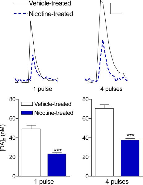
The present results show that long-term nicotine treatment significantly decreased dopamine release in rat NAcc shell, in agreement with our previous studies in monkeys (Perez et al. 2012). Single pulse and low frequency stimulation are commonly used to mimic tonic signaling while stimulus trains may be more closely associated with burst activity related to reward (Grace 2000). Therefore, release was measured using a single pulse, as well as a four-pulse stimulus train at 30 Hz as previous studies have shown that differential results may arise with varying firing frequency (Exley et al. 2008, Exley et al. 2011, Perez et al. 2008a, Rice & Cragg 2004, Zhang & Sulzer 2004, Zhang et al. 2009a). Both single pulse and four pulse-evoked dopamine release were reduced by ~50% compared to the vehicle-treated group (Fig. 1).
Fig. 1.
Long-term nicotine treatment decreases single pulse and 4 pulse-stimulated dopamine release in rat NAcc shell. Dopamine release [DA]o was measured in the dorsal portion of the accumbens shell in response to a single pulse or a four-pulse stimulus train delivered at 30 Hz. Representative traces of [DA]o are shown for vehicle- and nicotine-treated animals. Scale bar represents 10 nM and 0.5 s. Quantitative analyses show that long-term nicotine treatment significantly decreased [DA]o under both stimulation conditions. The values represent the mean ± SEM of 3-13 rats. Significance of difference from vehicle-treated rats, ***p < 0.001.
nAChR-mediated dopamine release in rat NAcc is primarily mediated via α6β2* nAChRs
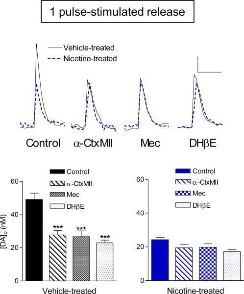
We next assessed the effect of nAChR antagonists on dopamine release in vehicle- and nicotine-treated rats. We first examined the nAChR subtypes that mediate dopamine release in vehicle-treated rats as previous studies have only been done in mouse and monkey NAcc (Exley et al. 2008, Exley et al. 2011, Perez et al. 2012). In vehicle-treated rats, the α6β2* nAChR antagonist α-conotoxinMII (α-CtxMII) decreased single pulse-stimulated dopamine release by ~45% (Fig. 2). No further decrease was observed upon application of the general nAChR antagonist mecamylamine (Mec) or the β2* nAChR antagonist dihydro-β-erythroidine (DHβE) (Fig. 2). These data show that α6β2* nAChRs regulate nAChR-mediated dopamine release in NAcc, consistent with work in mice and monkeys (Exley et al. 2008, Perez et al. 2012, Exley et al. 2011).
Fig. 2.
Long-term nicotine treatment abolishes the nAChR antagonist-induced decrease on single pulse-stimulated dopamine release. Dopamine release [DA]o was measured in the dorsal portion of the accumbens shell in response to single pulse electrical stimulation. Representative traces of [DA]o in the absence (control) and presence of the α6β2* antagonist α-CtxMII (100 nM), the general nAChR blocker Mec (100 μM), and the β2* nAChR antagonist DHβE (100 nM) are shown for vehicle- and nicotine-treated animals. Scale bar represents 10 nM and 0.5 s. Quantitative analyses show that α-CtxMII, Mec, and DHβE similarly affect single pulse-stimulated release in vehicle-treated animals, indicating that nAChR-modulated [DA]o occurs through α6β2* nAChRs. Single pulse-stimulated release was not further decreased in the presence of any of the nAChR antagonists after nicotine treatment. The values represent the mean ± SEM of 3-13 rats. Significance of difference from control release, ***p < 0.001.
In nicotine-treated rats, there was a significant 50% decrease in control single pulse-stimulated dopamine release compared to the vehicle-treated group (Fig. 2). However, in contrast to the results in vehicle-treated rats, exposure of the slices to α-CtxMII, Mec or DHβE did not significantly decrease single pulse-stimulated release (Fig. 2). These data suggest that long-term nicotine treatment primarily affects α6β2* nAChR-regulated dopamine release in rat accumbens.
Long-term nicotine treatment increases α4(non α6)β2* nAChRs and decreases α 6β2* nAChR expression
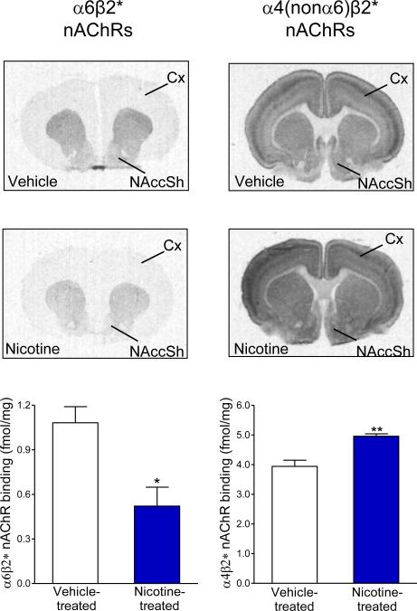
To investigate how the current long-term nicotine treatment modulated nAChR subtype expression in the NAcc, we measured α4β2* and α6β2* nAChRs. α4β2* nAChR binding was assessed using 125I-epibatidine autoradiography in the presence of the α6β2* nAChR antagonist α-CtxMII (Fig. 3). α4(nonα6)β2* nAChRs were significantly increased (p < 0.01) in the NAcc shell of nicotine-treated animals. We also assessed changes in α6β2* nAChRs using 125I-α-CtxMII binding (Fig. 3). This receptor subtype was significantly decreased (p < 0.05) with nicotine treatment. Thus, the decline in dopamine release with long-term nicotine treatment could be linked to decreases in this receptor subtype.
Fig. 3.
Long-term nicotine treatment decreases α6β2* nAChR expression and increases α4(nonα6)β2* nAChRs in rat NAcc. Representative autoradiograms of α6β2* nAChR binding using 125I-α-CtxMII and α4(nonα6)β2* nAChR binding using 125I-Epibatidine binding in the presence of α-CtxMII are shown for each treatment group. NAccSh, NAcc shell; Cx. Cortex. Quantitative analyses show a significant decrease in α6β2* nAChRs with an increase in α4(nonα6)β2* nAChRs after long-term nicotine treatment. Each value represents the mean ± SEM of 4 animals per group. Significance of difference from vehicle-treated rats, **p < 0.01,*p < 0.05.
Bath-applied nicotine decreases single pulse-stimulated dopamine release in slices of vehicle- and nicotine-treated rats
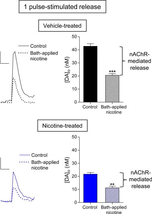
We next tested whether dopamine release was still sensitive to agonists after chronic nicotine treatment. To determine this, the exogenous agonist nicotine (300 nM) was bath-applied to slices from vehicle- and nicotine-treated animals and the effects on electrically-evoked dopamine release recorded. Bath-applied nicotine significantly decreased (p < 0.001) single pulse-stimulated dopamine release by ~52% in vehicle-treated rats (Fig. 4), in agreement with previous findings in rodents (Exley et al. 2008, Rice & Cragg 2004, Zhang & Sulzer 2004, Zhang et al. 2009b). A significant reduction (p < 0.01) in dopamine release (~49%) was also observed in slices from nicotine-treated rats (Fig. 4). These data indicate that the residual nAChRs are still functional and modulate dopamine release.
Fig. 4.
Decrease in single pulse-stimulated dopamine release in slices of vehicle- and nicotine-treated rats with bath-applied nicotine. Dopamine release [DA]o was determined in brain slices from vehicle-treated (top panel) and nicotine-treated (bottom panel) animals in the absence (control) and presence of bath-applied nicotine (300 nM), as shown in the representative traces. Scale bar represents 10 nM and 0.5 s. Quantitative analyses show that single pulse-evoked [DA]o was significantly decreased in the presence of bath-applied nicotine in both vehicle- and nicotine-treated animals, indicating that nAChRs in NAcc are responsive after long-term nicotine treatment. The values represent the mean ± SEM of 3-10 rats. Significance of difference from control, ***p < 0.001, **p < 0.01.
Acetylcholinesterase inhibition leads to similar effects on single pulse-stimulated release as bath-applied nicotine
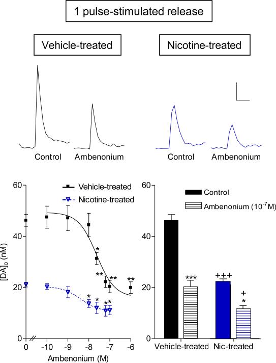
To further investigate the effect of agonists, we tested the acetylcholinesterase inhibitor ambenonium, which increases endogenous acetylcholine levels. The results show that ambenonium (0.1-1000 nM) attenuated dopamine release in slices of vehicle-treated animals up to 56% (p < 0.001), consistent with earlier work (Exley et al. 2008, Zhang et al. 2004). Acetylcholinesterase inhibition also affected release in nicotine-treated rats with a maximal 47% decline (p < 0.05). Dose-response curves show that ambenonium attenuated single-pulse stimulated dopamine release in a concentration-dependent manner in both treatment groups (Fig. 5). Notably, a significantly lower (p < 0.001) dose of ambenonium was required to induce a half-maximal inhibition of dopamine release in slices from nicotine-treated (IC50 = 4.81 ± 2.04 nM) versus vehicle-treated animals (IC50 = 24.1 ± 0.73 nM).
Fig. 5.
Decrease in single pulse-stimulated dopamine release in slices of vehicle- and nicotine-treated rats with the acetylcholinesterase inhibitor ambenonium. Dopamine release [DA]o was determined in the absence (control) and presence of ambenonium (0.1-1000 nM). Representative traces show that ambenonium decreases single pulse-stimulated [DA]o in slices from vehicle-and nicotine-treated rats. Scale bar represents 10 nM and 0.5 s. Dose-response curves are shown for vehicle- and nicotine-treated animals (Curve fits were sigmoidal with R = 0.8-0.9). Quantitative analyses show that ambenonium decreases release by approximately 50% at the maximal effective dose in both treatment groups (1000 nM and 100 nM for vehicle- and nicotine-treated respectively). The values represent the mean ± SEM of 3-4 rats. Significance of difference from control (zero ambenonium), ***p < 0.001, **p < 0.01, *p < 0.05. Significance of difference from vehicle-treated rats under each given condition, +++p < 0.001, +p < 0.05.
Long-term nicotine treatment does not affect dopamine transporter expression or dopamine uptake
Dopamine transporter levels were measured to determine whether the decline in release observed with long-term nicotine treatment might be due to a reduction in neurotransmitter transport. No differences in dopamine transporter levels were found between vehicle- (1.86 ± 0.10 fmol/mg tissue) and nicotine-treated animals (1.75 ± 0.05 fmol/mg tissue).
We also determined whether long-term nicotine treatment modified the kinetic constants describing dopamine reuptake following release. The decaying portion of each dopamine peak was fitted to a one-phase exponential decay at points where the amount of release matched in vehicle- and nicotine-treated animals as previously described (Cragg et al. 2001, John et al. 2006, Wightman & Zimmerman 1990, Yorgason et al. 2011). Nicotine treatment did not modify dopamine uptake rate constants (5.12 ± 0.26 and 4.46 ± 0.24 s−1 for vehicle- and nicotine-treated, respectively). Similarly, neither α-CtxMII, Mec, DHβE or frequency of stimulation had a significant effect on uptake rates in either treatment group (data not shown).
Effect of nAChR drugs and ambenonium on four pulse-stimulated dopamine release in slices from vehicle- and nicotine-treated animals
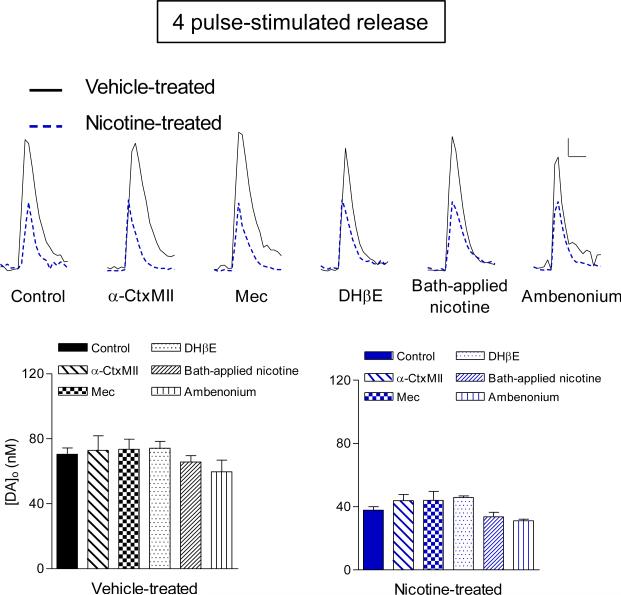
We also evaluated how long-term nicotine treatment altered dopamine release evoked via a four-pulse stimulus train at 30 Hz. Neither nAChR blockade, bath-applied nicotine nor ambenonium modified four pulse-stimulated release in vehicle-treated rats (Fig. 6). These findings agree with previous studies showing that nAChR antagonism or desensitization facilitates or does not change electrically-evoked dopamine release stimulated by high-frequencies (Exley & Cragg 2008, Exley et al. 2011, Perez et al. 2008a, Zhang & Sulzer 2004, Zhang et al. 2009a).
Fig. 6.
Effect of nAChR drugs and ambenonium on 4 pulse-stimulated dopamine release in slices from vehicle- and nicotine-treated animals. Dopamine release [DA]o was evoked via four-pulse stimulus train delivered at 30 Hz. Representative release signals in the absence (control) and presence of α-CtxMII (100nM), Mec (100μM), DHβE (100nM) and ambenonium (100nM) are shown for each treatment group. Scale bar represents 10 nM and 0.5 s. Quantitative analyses show that none of the drugs affect dopamine release in either vehicle- or nicotine-treated rat NAcc. The values represent the mean ± SEM of 3-15 rats.
Discussion
Enhanced dopamine release within the NAcc is a key event in nicotine reinforcement and reward (Balfour 2009, Corrigall et al. 1992, De Biasi & Dani 2011). β2, α4 and α6 nAChR subunits expressed in the mesolimbic dopaminergic pathway are involved in these functional and behavioral effects of nicotine, with extensive studies providing evidence for a role for α4β2* and α6β2* nAChRs (Changeux 2010, Tapper et al. 2004, Brunzell 2012, Epping-Jordan et al. 1999, Picciotto et al. 1998, Pons et al. 2008). Interestingly, converging work suggests that the α6α4β2* nAChR may be the subtype which plays a key regulatory role in endogenous dopamine neurotransmission both in the VTA and NAcc (Exley et al. 2011, Liu et al. 2012, Zhao-Shea et al. 2011), thus reconciling findings that suggest that only the α4β2* or α6β2* nAChR populations were involved. In view of the importance of nAChRs in regulating dopamine transmission and nicotine dependence, we initiated the present studies in slices containing the nucleus accumbens to further understand how long-term nicotine treatment modulated dopamine release.
Cyclic voltammetry studies showed that long-term nicotine treatment (2-3 months) decreased electrically-evoked dopamine release in the rat NAcc shell as it had in the monkeys (Perez et al. 2012). Thus, long-term nicotine exposure decreases dopamine release in response to both single and four pulse stimulation across species. To understand the nAChR subtypes that underlie the reduction in dopamine release, we tested the effect of the α6β2* nAChR blocker α-conotoxinMII, the β2*-selective antagonist dihydro-β-erythroidine and the general nAChR antagonist mecamylamine. The results in vehicle-treated rat NAcc show that similar declines were observed in single-pulse stimulated dopamine release with all the blockers. These data indicate that the α6β2* nAChR is the primary regulator of nAChR-mediated dopamine release, consistent with previous studies in mice and monkeys (Exley et al. 2008, Perez et al. 2012). The present data also showed a loss in nAChR-mediated dopamine release in nicotine-treated animals suggesting that alterations in nAChR expression and function contribute to the decrease in overall dopamine release, with the remaining release possibly reflecting mechanisms governed by direct electrically-evoked depolarization unaffected by nicotine treatment. Since the α6β2* nAChR is the major subtype that controls release in NAcc, function mediated by this nAChR subtype would thus be affected by long-term nicotine treatment.
As an alternate, possibly more direct approach to evaluate the effect of the current nicotine treatment regimen on nAChR levels, we did receptor autoradiography. A ~50% decrease was observed in NAcc α6β2* nAChR binding, consistent with the decline in dopamine release. The question remains which α6β2* nAChR subtype might be involved, that is, the α6α4β2* or α6β2* receptors. Previous work had shown that α6α4β2* nAChRs were decreased in rodent striatum after long-term nicotine treatment, with some increase in the α6β2* subtype (Perez et al. 2008a). These findings suggest that the decline in nAChR-modulated dopamine release with nicotine treatment may be partly due to a loss of α6α4β2* nAChRs, the primary regulator of nAChR-mediated release in NAcc. As well, the increase in α4(nonα6)β2* nAChRs, which are predominantly located post-synaptically on inhibitory GABAergic terminals and interneurons (Quik & Wonnacott 2011) could also play a role. It should be noted however that there are limitations of the binding assays when comparing the effects of nicotine on α4β2* and α6β2* nAChRs. Unlike epibatidine which binds to desensitized receptors and may recognize immature complexes, α-conotoxin may only bind to mature α6β2* receptors. Moreover, epibatidine is an agonist whereas α-conotoxinMII is an antagonist. Thus caution is warranted with respect to interpretation of the nAChR autoradiography.
An important issue was whether the evoked dopamine release component remaining after long-term nicotine dosing was sensitive to the desensitizing effects of nicotine on dopamine release, that is, was the residual release still regulated by nAChRs. To test this, we used a nicotine concentration of 300 nM, which is similar to that reported in the plasma of smokers (Jarvik et al. 2000). Dopamine release was still responsive to bath-applied nicotine, suggesting that release was still regulated by nAChRs in NAcc of nicotine-treated rats. Similar results were obtained with the acetylcholinesterase inhibitor ambenonium, which sustains enhanced endogenous acetylcholine levels, hence leading to desensitization. These combined findings support the idea that although nAChR desensitization most likely contributes to the decrease in nAChR-mediated release, there are receptors that are still functional after long-term nicotine dosing. This conclusion would agree with recent work showing that nicotine desensitizes α6β2* nAChRs to a lesser extent than other nAChR subtypes such that only 25% of their function is lost (Grady et al. 2012, Kuryatov & Lindstrom 2011). As a result, these receptors could still partially respond to the desensitizing effects of prolonged agonist exposure. Such an effect may provide a rationale as to why cigarettes are still rewarding to and craved by chronic smokers.
The data showing that dopamine release evoked by a train of pulses (4 pulses) at 30Hz was similar in the absence or presence of agonists and antagonists is consistent with previous studies (Meyer et al. 2008, Perez et al. 2009, Zhang & Sulzer 2004, Zhang et al. 2009a, Zhang et al. 2009b). This lack of effect of nAChR antagonism or desensitization on burst-induced release is due to a relief of short-term depression. Tonically active cholinergic interneurons provide a constant source of acetylcholine in the striatum and nucleus accumbens both in vivo and in vitro (Exley & Cragg 2008, Rice et al. 2011). This endogenous acetylcholine promotes dopamine release by interacting at nAChRs (Cragg 2003, De Biasi & Dani 2011). With a burst of action potentials, however, the initial enhanced dopamine release limits release with subsequent pulses – a phenomenon known as short-term depression (Cragg 2003). In the presence of nAChR antagonists or with desensitization, dopamine release is partially inhibited so that the first pulse induces a smaller amount of dopamine release due to receptor blockade. As a result, there is less short-term depression. Subsequent pulses within a burst cause more dopamine release such that overall release with burst firing is similar or enhanced in the presence of antagonists or with desensitization. These effects of nicotinic receptor blockade/desensitization may mimic the pause in cholinergic interneuron activity observed in vivo upon the presentation of reward-related events while dopamine neurons switch into bursting mode (Exley & Cragg 2008, Rice et al. 2011).
The finding that agonists but not antagonists affect dopamine release in the NAcc of nicotine-treated rats may have two implications. First, nicotine treatment may modify acetylcholine tone. Since antagonists generally act by blocking the action of endogenous acetylcholine at nAChRs, a possible explanation for their lack of effect may be that long-term nicotine treatment reduces acetylcholine levels. This idea is consistent with findings showing that antagonist-dependent changes in electrically-evoked release require an interaction of endogenous acetylcholine with nAChRs (Exley et al. 2008, Zhang & Sulzer 2004, Zhou et al. 2001). Recent work also shows that endogenous acetylcholine release arising from the synchronized activity of cholinergic interneurons directly influences dopamine release regardless of the extent of dopaminergic activity (Cachope et al. 2012, Threlfell et al. 2012). Alternately, the ability of desensitizing concentrations of nicotine or acetylcholinesterase inhibition to further decrease dopamine release may be independent of α6β2* and α4β2* nAChRs. For instance, nAChR agonists may stimulate α7 nAChRs on glutamatergic terminals and indirectly modulate release from dopaminergic terminals. Studies to determine whether such a mechanisms contribute to the effects of agonists after long-term nicotine treatment remain to be done.
A question that arises concerns reconciliation of the observed decrease in nAChR receptor number and function following prolonged nicotine administration via drinking water with in vivo studies showing a sensitized response to nicotine after chronic nicotine exposure. One factor that may influence these differential effects of nicotine on dopamine signaling is route of administration. Indeed, studies show that sensitization is observed following daily nicotine injections but not chronic nicotine infusions (Benwell & Balfour 1997). Our drinking water model may replicate the sustained nicotine levels experienced with smoking and provide information about the functional effects of this long-term exposure. Conversely, measurements during an in vivo injection after nicotine exposure may better mimic the changes in dopamine release that occur during the nicotine peaks deemed essential for reward. An additional factor is that the brain slice preparation used in our studies is an isolated system devoid of the circuitry normally influencing the nucleus accumbens. Thus, it does not yield information on how changes in the expression and function of α6β2* nAChRs in the VTA contribute to dopamine release in the accumbens shell in vivo after long-term nicotine exposure. Therefore, bath-application of nicotine may not fully replicate the in vivo effects of an acute nicotine exposure. Nevertheless, nAChRs at the terminal level are known to play an important role in directly modulating dopamine release and it is important to determine how nicotine exposure modifies their function as such local alterations may directly or indirectly affect signaling in the entire reward pathway.
In summary, the present results suggest that long-term nicotine decreases NAcc dopamine release via a downregulation of α6β2* nAChR levels and a decline in their function, possibly due to reduced extracellular acetylcholine levels. These findings, coupled with observations that the smoking habit is thought to arise from the need to preserve nicotine-induced changes (Paolini & De Biasi 2011), support two possible therapeutic strategies to treat nicotine withdrawal. First, therapies that enhance the action of acetylcholine may help alleviate the urge to smoke by mimicking the functional effects of nicotine. Indeed, the use of acetylcholinesterase inhibitors is being explored as a potential smoking cessation therapy, with the advantage that such drugs could also reverse withdrawal-induced cognitive impairments (Hopkins et al. 2012, Sofuoglu & Mooney 2009). Second, our results suggest that α6β2* nAChR agonists may represent a useful approach to combat nicotine dependence because of their prominent modulatory control of accumbal dopaminergic signaling.
Acknowledgements
The authors would like to thank Jason Ly for excellent technical assistance. This work was supported by the National Institutes of Health [Grants NS 59910, GM103801 and GM48677]; and the California Tobacco Related Disease Research Program [Grant 17RT-0119]. The authors declare no conflict of interest. MQ and XAP designed the study and wrote the manuscript. XAP performed all experiments and analyzed the data. JMM contributed the α-conotoxinMII and critically discussed the manuscript. All authors approved the submitted version of the manuscript.
Abbreviations used
- nAChRs
nicotinic acetylcholine receptors
- α-CtxMII
α-conotoxinMII
- Mec
Mecamylamine
- DHβE
dihydro-β-erythroidine
- RTI-121
3β-(4-iodophenyl)tropane-2β-carboxylic acid
- *
the asterisk indicates the possible presence of other nicotinic subunits in the receptor complex, with α4β2* nAChRs including α4β2 and α4α5β2 subtypes, and α6β2* including α6α4β2β3, α6β2β3 and α6β2 subtypes
References
- Balfour DJ. The neuronal pathways mediating the behavioral and addictive properties of nicotine. Handb Exp Pharmacol. 2009:209–233. doi: 10.1007/978-3-540-69248-5_8. [DOI] [PubMed] [Google Scholar]
- Benowitz NL. Nicotine addiction. N Engl J Med. 2010;362:2295–2303. doi: 10.1056/NEJMra0809890. [DOI] [PMC free article] [PubMed] [Google Scholar]
- Benwell ME, Balfour DJ. Regional variation in the effects of nicotine on catecholamine overflow in rat brain. Eur J Pharmacol. 1997;325:13–20. doi: 10.1016/s0014-2999(97)00101-5. [DOI] [PubMed] [Google Scholar]
- Brunzell DH. Preclinical Evidence That Activation of Mesolimbic Alpha 6 Subunit Containing Nicotinic Acetylcholine Receptors Supports Nicotine Addiction Phenotype. Nicotine Tob Res. 2012;14:1258–1269. doi: 10.1093/ntr/nts089. [DOI] [PMC free article] [PubMed] [Google Scholar]
- Cachope R, Mateo Y, Mathur BN, Irving J, Wang HL, Morales M, Lovinger DM, Cheer JF. Selective activation of cholinergic interneurons enhances accumbal phasic dopamine release: setting the tone for reward processing. Cell Rep. 2012;2:33–41. doi: 10.1016/j.celrep.2012.05.011. [DOI] [PMC free article] [PubMed] [Google Scholar]
- Changeux JP. Nicotine addiction and nicotinic receptors: lessons from genetically modified mice. Nat Rev Neurosci. 2010;11:389–401. doi: 10.1038/nrn2849. [DOI] [PubMed] [Google Scholar]
- Corrigall WA, Franklin KB, Coen KM, Clarke PB. The mesolimbic dopaminergic system is implicated in the reinforcing effects of nicotine. Psychopharmacology (Berl) 1992;107:285–289. doi: 10.1007/BF02245149. [DOI] [PubMed] [Google Scholar]
- Cragg SJ. Variable dopamine release probability and short-term plasticity between functional domains of the primate striatum. J Neurosci. 2003;23:4378–4385. doi: 10.1523/JNEUROSCI.23-10-04378.2003. [DOI] [PMC free article] [PubMed] [Google Scholar]
- Cragg SJ, Nicholson C, Kume-Kick J, Tao L, Rice ME. Dopamine-mediated volume transmission in midbrain is regulated by distinct extracellular geometry and uptake. J Neurophysiol. 2001;85:1761–1771. doi: 10.1152/jn.2001.85.4.1761. [DOI] [PubMed] [Google Scholar]
- De Biasi M, Dani JA. Reward, addiction, withdrawal to nicotine. Annu Rev Neurosci. 2011;34:105–130. doi: 10.1146/annurev-neuro-061010-113734. [DOI] [PMC free article] [PubMed] [Google Scholar]
- Dhelaria RK, Friderici J, Wu K, Gupta E, Khan C, Rothberg MB. Effectiveness of varenicline for smoking cessation at 2 urban academic health centers. Eur J Intern Med. 2012;23:461–464. doi: 10.1016/j.ejim.2012.03.017. [DOI] [PubMed] [Google Scholar]
- Di Chiara G, Bassareo V, Fenu S, et al. Dopamine and drug addiction: the nucleus accumbens shell connection. Neuropharmacology. 2004;47(Suppl 1):227–241. doi: 10.1016/j.neuropharm.2004.06.032. [DOI] [PubMed] [Google Scholar]
- Epping-Jordan MP, Picciotto MR, Changeux JP, Pich EM. Assessment of nicotinic acetylcholine receptor subunit contributions to nicotine self-administration in mutant mice. Psychopharmacology (Berl) 1999;147:25–26. doi: 10.1007/s002130051135. [DOI] [PubMed] [Google Scholar]
- Exley R, Clements MA, Hartung H, McIntosh JM, Cragg SJ. Alpha6-containing nicotinic acetylcholine receptors dominate the nicotine control of dopamine neurotransmission in nucleus accumbens. Neuropsychopharmacology. 2008;33:2158–2166. doi: 10.1038/sj.npp.1301617. [DOI] [PubMed] [Google Scholar]
- Exley R, Cragg SJ. Presynaptic nicotinic receptors: a dynamic and diverse cholinergic filter of striatal dopamine neurotransmission. Br J Pharmacol. 2008;153(Suppl 1):S283–297. doi: 10.1038/sj.bjp.0707510. [DOI] [PMC free article] [PubMed] [Google Scholar]
- Exley R, Maubourguet N, David V, et al. Distinct contributions of nicotinic acetylcholine receptor subunit alpha4 and subunit alpha6 to the reinforcing effects of nicotine. Proc Natl Acad Sci U S A. 2011;108:7577–7582. doi: 10.1073/pnas.1103000108. [DOI] [PMC free article] [PubMed] [Google Scholar]
- Gotti C, Guiducci S, Tedesco V, et al. Nicotinic acetylcholine receptors in the mesolimbic pathway: primary role of ventral tegmental area alpha6beta2* receptors in mediating systemic nicotine effects on dopamine release, locomotion, and reinforcement. J Neurosci. 2010;30:5311–5325. doi: 10.1523/JNEUROSCI.5095-09.2010. [DOI] [PMC free article] [PubMed] [Google Scholar]
- Grace AA. The tonic/phasic model of dopamine system regulation and its implications for understanding alcohol and psychostimulant craving. Addiction. 2000;95(Suppl 2):S119–128. doi: 10.1080/09652140050111690. [DOI] [PubMed] [Google Scholar]
- Grady SR, Wageman CR, Patzlaff NE, Marks MJ. Low concentrations of nicotine differentially desensitize nicotinic acetylcholine receptors that include alpha5 or alpha6 subunits and that mediate synaptosomal neurotransmitter release. Neuropharmacology. 2012;62:1935–1943. doi: 10.1016/j.neuropharm.2011.12.026. [DOI] [PMC free article] [PubMed] [Google Scholar]
- Hopkins TJ, Rupprecht LE, Hayes MR, Blendy JA, Schmidt HD. Galantamine, an Acetylcholinesterase Inhibitor and Positive Allosteric Modulator of Nicotinic Acetylcholine Receptors, Attenuates Nicotine Taking and Seeking in Rats. Neuropsychopharmacology. 2012;37:2310–2321. doi: 10.1038/npp.2012.83. [DOI] [PMC free article] [PubMed] [Google Scholar]
- Jackson KJ, McIntosh JM, Brunzell DH, Sanjakdar SS, Damaj MI. The role of alpha6-containing nicotinic acetylcholine receptors in nicotine reward and withdrawal. J Pharmacol Exp Ther. 2009;331:547–554. doi: 10.1124/jpet.109.155457. [DOI] [PMC free article] [PubMed] [Google Scholar]
- Jarvik ME, Madsen DC, Olmstead RE, Iwamoto-Schaap PN, Elins JL, Benowitz NL. Nicotine blood levels and subjective craving for cigarettes. Pharmacol Biochem Behav. 2000;66:553–558. doi: 10.1016/s0091-3057(00)00261-6. [DOI] [PubMed] [Google Scholar]
- John CE, Budygin EA, Mateo Y, Jones SR. Neurochemical characterization of the release and uptake of dopamine in ventral tegmental area and serotonin in substantia nigra of the mouse. J Neurochem. 2006;96:267–282. doi: 10.1111/j.1471-4159.2005.03557.x. [DOI] [PubMed] [Google Scholar]
- Keating GM, Lyseng-Williamson KA. Varenicline: a pharmacoeconomic review of its use as an aid to smoking cessation. Pharmacoeconomics. 2010;28:231–254. doi: 10.2165/11204380-000000000-00000. [DOI] [PubMed] [Google Scholar]
- Kuryatov A, Lindstrom J. Expression of functional human alpha6beta2beta3* acetylcholine receptors in Xenopus laevis oocytes achieved through subunit chimeras and concatamers. Mol Pharmacol. 2011;79:126–140. doi: 10.1124/mol.110.066159. [DOI] [PMC free article] [PubMed] [Google Scholar]
- Liu L, Zhao-Shea R, McIntosh JM, Gardner PD, Tapper AR. Nicotine persistently activates ventral tegmental area dopaminergic neurons via nicotinic acetylcholine receptors containing alpha4 and alpha6 subunits. Mol Pharmacol. 2012;81:541–548. doi: 10.1124/mol.111.076661. [DOI] [PMC free article] [PubMed] [Google Scholar]
- Matta SG, Balfour DJ, Benowitz NL, et al. Guidelines on nicotine dose selection for in vivo research. Psychopharmacology (Berl) 2007;190:269–319. doi: 10.1007/s00213-006-0441-0. [DOI] [PubMed] [Google Scholar]
- Meyer EL, Yoshikami D, McIntosh JM. The Neuronal Nicotinic Acetylcholine Receptors alpha4 and alpha6 Differentially Modulate Dopamine Release in Mouse Striatal Slices. J Neurochem. 2008 doi: 10.1111/j.1471-4159.2008.05266.x. doi:10.1111/j.1471-4159.2008.05266.x. [DOI] [PMC free article] [PubMed] [Google Scholar]
- Mills EJ, Wu P, Lockhart I, Thorlund K, Puhan M, Ebbert JO. Comparisons of high-dose and combination nicotine replacement therapy, varenicline, and bupropion for smoking cessation: A systematic review and multiple treatment meta-analysis. Ann Med. 2012 doi: 10.3109/07853890.2012.705016. [DOI] [PubMed] [Google Scholar]
- Paolini M, De Biasi M. Mechanistic insights into nicotine withdrawal. Biochem Pharmacol. 2011;82:996–1007. doi: 10.1016/j.bcp.2011.07.075. [DOI] [PMC free article] [PubMed] [Google Scholar]
- Perez XA, Bordia T, McIntosh JM, Grady SR, Quik M. Long-term nicotine treatment differentially regulates striatal alpha6alpha4beta2* and alpha6(nonalpha4)beta2* nAChR expression and function. Mol Pharmacol. 2008a;74:844–853. doi: 10.1124/mol.108.048843. [DOI] [PMC free article] [PubMed] [Google Scholar]
- Perez XA, Bordia T, McIntosh JM, Quik M. alpha6ss2* and alpha4ss2* nicotinic receptors both regulate dopamine signaling with increased nigrostriatal damage: relevance to Parkinson's disease. Mol Pharmacol. 2010;78:971–980. doi: 10.1124/mol.110.067561. [DOI] [PMC free article] [PubMed] [Google Scholar]
- Perez XA, Ly J, McIntosh JM, Quik M. Long-term nicotine exposure depresses dopamine release in nonhuman primate nucleus accumbens. J Pharmacol Exp Ther. 2012;342:335–344. doi: 10.1124/jpet.112.194084. [DOI] [PMC free article] [PubMed] [Google Scholar]
- Perez XA, O'Leary KT, Parameswaran N, McIntosh JM, Quik M. Prominent role of alpha3/alpha6beta2* nAChRs in regulating evoked dopamine release in primate putamen: effect of long-term nicotine treatment. Mol Pharmacol. 2009;75:938–946. doi: 10.1124/mol.108.053801. [DOI] [PMC free article] [PubMed] [Google Scholar]
- Perez XA, Parameswaran N, Huang LZ, O'Leary KT, Quik M. Pre-synaptic dopaminergic compensation after moderate nigrostriatal damage in non-human primates. J Neurochem. 2008b;105:1861–1872. doi: 10.1111/j.1471-4159.2008.05268.x. [DOI] [PMC free article] [PubMed] [Google Scholar]
- Picciotto MR, Zoli M, Rimondini R, Lena C, Marubio LM, Pich EM, Fuxe K, Changeux JP. Acetylcholine receptors containing the beta2 subunit are involved in the reinforcing properties of nicotine. Nature. 1998;391:173–177. doi: 10.1038/34413. [DOI] [PubMed] [Google Scholar]
- Pons S, Fattore L, Cossu G, Tolu S, Porcu E, McIntosh JM, Changeux JP, Maskos U, Fratta W. Crucial role of alpha4 and alpha6 nicotinic acetylcholine receptor subunits from ventral tegmental area in systemic nicotine self-administration. J Neurosci. 2008;28:12318–12327. doi: 10.1523/JNEUROSCI.3918-08.2008. [DOI] [PMC free article] [PubMed] [Google Scholar]
- Quik M, Polonskaya Y, Gillespie A, G KL, Langston JW. Differential alterations in nicotinic receptor alpha6 and beta3 subunit messenger RNAs in monkey substantia nigra after nigrostriatal degeneration. Neuroscience. 2000;100:63–72. doi: 10.1016/s0306-4522(00)00244-x. [DOI] [PubMed] [Google Scholar]
- Quik M, Polonskaya Y, Kulak JM, McIntosh JM. Vulnerability of 125I-alpha-conotoxin MII binding sites to nigrostriatal damage in monkey. J Neurosci. 2001;21:5494–5500. doi: 10.1523/JNEUROSCI.21-15-05494.2001. [DOI] [PMC free article] [PubMed] [Google Scholar]
- Quik M, Wonnacott S. alpha6beta2* and alpha4beta2* nicotinic acetylcholine receptors as drug targets for Parkinson's disease. Pharmacol Rev. 2011;63:938–966. doi: 10.1124/pr.110.003269. [DOI] [PMC free article] [PubMed] [Google Scholar]
- Rice ME, Cragg SJ. Nicotine amplifies reward-related dopamine signals in striatum. Nat Neurosci. 2004;7:583–584. doi: 10.1038/nn1244. [DOI] [PubMed] [Google Scholar]
- Rice ME, Patel JC, Cragg SJ. Dopamine release in the basal ganglia. Neuroscience. 2011;198:112–137. doi: 10.1016/j.neuroscience.2011.08.066. [DOI] [PMC free article] [PubMed] [Google Scholar]
- Sellings LH, Baharnouri G, McQuade LE, Clarke PB. Rewarding and aversive effects of nicotine are segregated within the nucleus accumbens. Eur J Neurosci. 2008;28:342–352. doi: 10.1111/j.1460-9568.2008.06341.x. [DOI] [PubMed] [Google Scholar]
- Sofuoglu M, Mooney M. Cholinergic functioning in stimulant addiction: implications for medications development. CNS Drugs. 2009;23:939–952. doi: 10.2165/11310920-000000000-00000. [DOI] [PMC free article] [PubMed] [Google Scholar]
- Tapper AR, McKinney SL, Nashmi R, et al. Nicotine activation of alpha4* receptors: sufficient for reward, tolerance, and sensitization. Science. 2004;306:1029–1032. doi: 10.1126/science.1099420. [DOI] [PubMed] [Google Scholar]
- Threlfell S, Lalic T, Platt NJ, Jennings KA, Deisseroth K, Cragg SJ. Striatal dopamine release is triggered by synchronized activity in cholinergic interneurons. Neuron. 2012;75:58–64. doi: 10.1016/j.neuron.2012.04.038. [DOI] [PubMed] [Google Scholar]
- Tuesta LM, Fowler CD, Kenny PJ. Recent advances in understanding nicotinic receptor signaling mechanisms that regulate drug self-administration behavior. Biochem Pharmacol. 2011;82:984–995. doi: 10.1016/j.bcp.2011.06.026. [DOI] [PMC free article] [PubMed] [Google Scholar]
- Wightman RM, Zimmerman JB. Control of dopamine extracellular concentration in rat striatum by impulse flow and uptake. Brain Res Brain Res Rev. 1990;15:135–144. doi: 10.1016/0165-0173(90)90015-g. [DOI] [PubMed] [Google Scholar]
- Wise RA. Roles for nigrostriatal--not just mesocorticolimbic--dopamine in reward and addiction. Trends Neurosci. 2009;32:517–524. doi: 10.1016/j.tins.2009.06.004. [DOI] [PMC free article] [PubMed] [Google Scholar]
- Yorgason JT, Espana RA, Jones SR. Demon Voltammetry and Analysis software: Analysis of cocaine-induced alterations in dopamine signaling using multiple kinetic measures. J Neurosci Methods. 2011 doi: 10.1016/j.jneumeth.2011.03.001. [DOI] [PMC free article] [PubMed] [Google Scholar]
- Zhang H, Sulzer D. Frequency-dependent modulation of dopamine release by nicotine. Nat Neurosci. 2004;7:581–582. doi: 10.1038/nn1243. [DOI] [PubMed] [Google Scholar]
- Zhang L, Dong Y, Doyon WM, Dani JA. Withdrawal from Chronic Nicotine Exposure Alters Dopamine Signaling Dynamics in the Nucleus Accumbens. Biol Psychiatry. 2011 doi: 10.1016/j.biopsych.2011.07.024. [DOI] [PMC free article] [PubMed] [Google Scholar]
- Zhang L, Doyon WM, Clark JJ, Phillips PE, Dani JA. Controls of tonic and phasic dopamine transmission in the dorsal and ventral striatum. Mol Pharmacol. 2009a;76:396–404. doi: 10.1124/mol.109.056317. [DOI] [PMC free article] [PubMed] [Google Scholar]
- Zhang L, Zhou FM, Dani JA. Cholinergic drugs for Alzheimer's disease enhance in vitro dopamine release. Mol Pharmacol. 2004;66:538–544. doi: 10.1124/mol.104.000299. [DOI] [PubMed] [Google Scholar]
- Zhang T, Zhang L, Liang Y, Siapas AG, Zhou FM, Dani JA. Dopamine signaling differences in the nucleus accumbens and dorsal striatum exploited by nicotine. J Neurosci. 2009b;29:4035–4043. doi: 10.1523/JNEUROSCI.0261-09.2009. [DOI] [PMC free article] [PubMed] [Google Scholar]
- Zhao-Shea R, Liu L, Soll LG, et al. Nicotine-mediated activation of dopaminergic neurons in distinct regions of the ventral tegmental area. Neuropsychopharmacology. 2011;36:1021–1032. doi: 10.1038/npp.2010.240. [DOI] [PMC free article] [PubMed] [Google Scholar]
- Zhou FM, Liang Y, Dani JA. Endogenous nicotinic cholinergic activity regulates dopamine release in the striatum. Nat Neurosci. 2001;4:1224–1229. doi: 10.1038/nn769. [DOI] [PubMed] [Google Scholar]
- Zhu SH, Lee M, Zhuang YL, Gamst A, Wolfson T. Interventions to increase smoking cessation at the population level: how much progress has been made in the last two decades? Tob Control. 2012;21:110–118. doi: 10.1136/tobaccocontrol-2011-050371. [DOI] [PMC free article] [PubMed] [Google Scholar]


