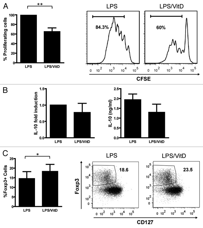
Figure 4. Skin DCs migrating out of VitD-injected skin induce the development of Foxp3+ Tregs. T cells induced by LPS- or LPS/VitD-primed skin crawl-out cells were analyzed for regulatory characteristics. (A) The suppressive capacity of induced T cells expressed as the proliferation of bystander T cells. This proliferation is depicted as the percentage of T cell proliferation of the LPS condition, which was set as a base line. (B) IL-10 fold induction in 24 h-supernatants of T cells restimulated by αCD3/αCD28. (C) Foxp3 expression by resting T cells was determined by flow cytometry. Results are representative of 5 (A, right panel), 6 (B, right panel) or 7 (C, right panel) or the mean ± SEM of 5 (A left panel), 6 (B, left panel) or 7 (C, left panel) independent experiments.*p < 0.05, **p < 0.01.

An official website of the United States government
Here's how you know
Official websites use .gov
A
.gov website belongs to an official
government organization in the United States.
Secure .gov websites use HTTPS
A lock (
) or https:// means you've safely
connected to the .gov website. Share sensitive
information only on official, secure websites.
